6 Neurons and neuroglia
Overview
Nerve cells, or neurons, are the structural and functional units of the nervous system. They generate and conduct electrical changes in the form of nerve impulses. They communicate chemically with other neurons at points of contact called synapses. Neuroglia (literally, ‘nerve glue’) is the connective tissue of the nervous system.
Neuroglial cells outnumber neurons by about five to one. They have important nutritive and supportive functions.
Neurons
Billions of neurons form a shell, or cortex, on the surface of the cerebral and cerebellar hemispheres. In this general context, nuclei are aggregates of neurons buried within the white matter.
In the central nervous system (CNS), almost all neurons are multipolar, their cell bodies or somas having multiple poles or angular points. At every pole but one, a dendrite emerges and divides repeatedly (Figure 6.1). On some neurons, the shafts of the dendrites are smooth. On others, the shafts show numerous short spines (Figure 6.2). The dendrites receive synaptic contacts from other neurons, from some on the spines and from others on the shafts.

Figure 6.1 Profiles of neurons from the brain. (1) Pyramidal cell, cerebral cortex. (2) Neuroendocrine cell, hypothalamus. (3) Spiny neuron, corpus striatum. (4) Basket cell, cerebellum. Neurons 1 and 3 show dendritic spines. A, axon; AC, axon collateral; D, dendrites.

Figure 6.2 Dendritic spines. This section is taken from the cerebellum, where the dendrites of the giant cells of Purkinje are studded with spines. In this field, three spines (S) are in receipt of synaptic contacts by axonic boutons (A). A fourth axon (top left) is synapsing on the shaft of the dendrite.
(From Pannese 1994, with permission of Thieme.)
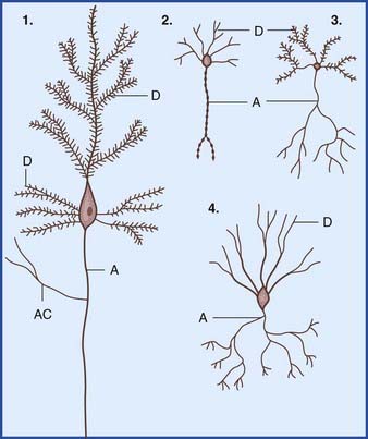
The remaining pole of the soma gives rise to the axon, which conducts nerve impulses. Most axons give off collateral branches (Figure 6.3). Terminal branches synapse on target neurons.

Figure 6.3 (A) Motor neuron in the anterior gray horn of the spinal cord. (B) Enlargement from (A). Myelin segments 1 and 2 occupy central nervous system white matter and have been laid down by an oligodendrocyte; a recurrent collateral branch of the axon originated from the node. Myelin segments 3 and 4 occupy peripheral nervous system and have been laid down by Schwann cells; the node at the transitional zone is bounded by an oligodendrocyte and a Schwann cell. (C) Neurofibrils (matted neurofilaments) are seen after staining with silver salts. (D) Nissl bodies (clumps of granular endoplasmic reticulum) are seen after staining with a cationic dye such as thionin.
Most synaptic contacts between neurons are either axodendritic or axosomatic. Axodendritic synapses are usually excitatory in their effect on target neurons, whereas most axosomatic synapses have an inhibitory effect.
Internal structure of neurons
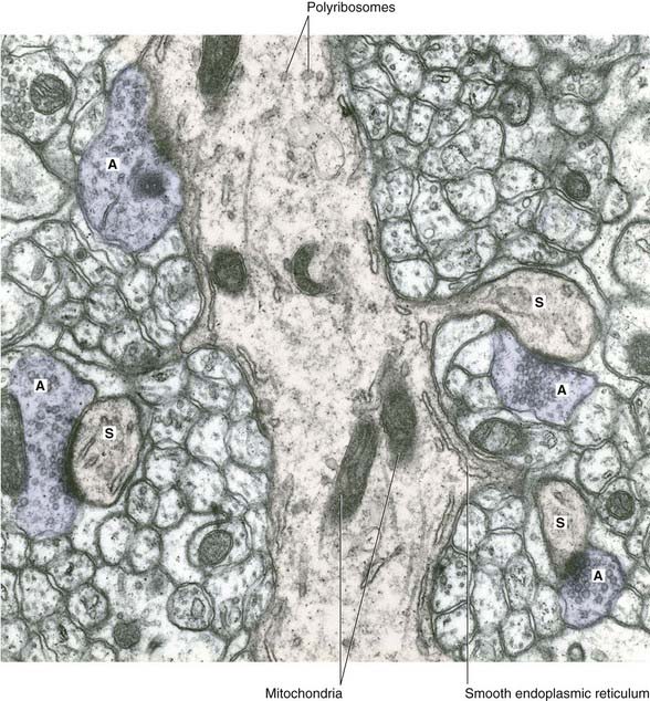
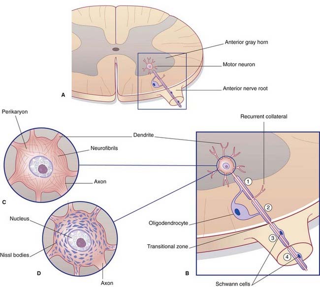
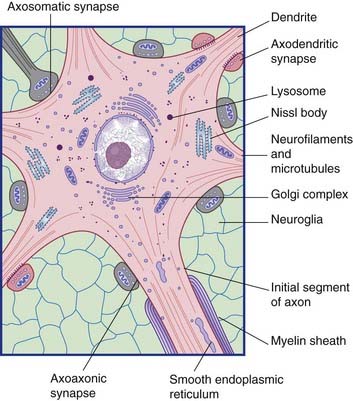
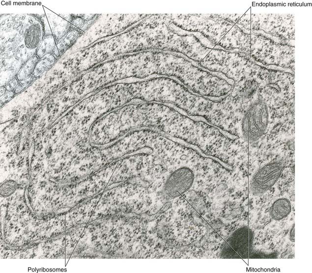
All parts of neurons are permeated by microtubules and neurofilaments (Figure 6.4). The soma contains the nucleus and the cytoplasm or perikaryon (Gr.’around the nucleus’). The perikaryon contains clumps of granular endoplasmic reticulum known as Nissl bodies (Figure 6.5), also Golgi complexes, free ribosomes, mitochondria, and smooth endoplasmic reticulum (Figure 6.4).

Figure 6.4 Ultrastructure of a motor neuron. Stems of five dendrites are included, also three excitatory synapses (red) and five inhibitory synapses.

Figure 6.5 Nissl substance in the soma of a motor neuron. The endoplasmic reticulum has a characteristic stacked arrangement. Polyribosomes are studded along the outer surface of the cisternae; many others lie free in the cytoplasm. (Note: Faint color tones have been added here and later for ease of identification.)
(From Pannese 1994, with permission of Thieme.)
Intracellular transport
Turnover of membranous and skeletal materials takes place in all cells. In neurons, fresh components are continuously synthesized in the soma and moved into the axon and dendrites by a process of anterograde transport. At the same time, worn-out materials are returned to the soma by retrograde transport for degradation in lysosomes (see also target recognition, later).
Anterograde transport is of two kinds: rapid and slow. Included in rapid transport (at a speed of 300–400 mm/day) are free elements such as synaptic vesicles, transmitter substances (or their precursor molecules), and mitochondria. Also included are lipid and protein molecules (including receptor proteins) for insertion into the plasma membrane. Included in slow transport (at 5–10 mm/day) are the skeletal elements, and soluble proteins including some of those involved in transmitter release at nerve endings. Microtubules seem to be largely constructed within the axon. They are exported from the soma in preassembled short sheaves that propel one another along the initial segment of the axon; further progress is mainly by a process of elongation (up to 1 mm apiece) performed by the addition of tubulin polymers at their distal ends, with some disassembly at their proximal ends. The bulk movement of neurofilaments slows down to almost zero distally; there, the filaments are refreshed by the insertion of filament polymers moving from the soma by slow transport.
Retrograde transport of worn-out mitochondria, SER, and plasma membrane (including receptors therein) is fairly rapid (150–200 mm/day). In addition to its function in waste disposal, retrograde transport is involved in target cell recognition. At synaptic contacts, axons constantly ‘nibble’ the plasma membrane of target neurons by means of endocytotic uptake of protein-containing signaling endosomes. These proteins are known as neurotrophins (‘neuron foods’). They are brought to the soma and incorporated into Golgi complexes there. In addition, uptake of target cell ‘marker’ molecules is important for cell recognition during development. It may also be necessary for viability later on, because adult neurons shrink and may even die if their axons are severed proximal to their first branches.
The longest-known neurotrophin is nerve growth factor, on which the developing peripheral sensory and autonomic systems are especially dependent. Adult brain neurons synthesize brain-derived neurotrophic factor (BDNF) in the soma and send it to their nerve endings by anterograde transport. Animal studies have shown that BDNF maintains the general health of neurons in terms of metabolic activity, impulse propagation, and synaptic transmission.
Transport mechanisms
Microtubules are the supporting structures for neuronal transport. Microtubule-binding proteins, in the form of ATPases, propel organelles and molecules along the outer surface of the microtubules. Distinct ATPases are used for anterograde and retrograde work. Retrograde transport of signaling endosomes is performed by the dynein ATPase. Failure of dynein performance has been linked to motor neuron disease, described in Chapter 16.
Some points of clinical relevance are highlighted in Clinical Panel 6.1.
Clinical Panel 6.1 Clinical relevance of neuronal transport
Tetanus
Wounds contaminated by soil or street dust may contain Clostridium tetani. The toxin produced by this organism binds to the plasma membrane of nerve endings, is taken up by endocytosis, and is carried to the spinal cord by retrograde transport. Other neurons upstream take in the toxin by endocytosis – notably Renshaw cells (Ch. 15), which normally exert a braking action on motor neurons through the release of an inhibitory transmitter substance, glycine. Tetanus toxin prevents the release of glycine. As a result, motor neurons go out of control, particularly those supplying the muscles of the face, jaws, and spine. These muscles exhibit prolonged, agonizing spasms. About half of the patients who show these classic signs of tetanus die of exhaustion within a few days. Tetanus is entirely preventable by appropriate and timely immunization.
Viruses and toxic metals
Retrograde axonal transport has been blamed for the passage of viruses from the nasopharynx to the central nervous system, also for the uptake of toxic metals such as lead and aluminum. Viruses, in particular, may be spread widely through the brain by means of retrograde transneuronal uptake.

Stay updated, free articles. Join our Telegram channel

Full access? Get Clinical Tree

