Normal EEG and Sleep: Infants to Adolescents


Normal EEG and Sleep: Infants to Adolescents


James J. Riviello Jr

Douglas R. Nordli Jr

Ernst Niedermeyer


GENERAL PRINCIPLES

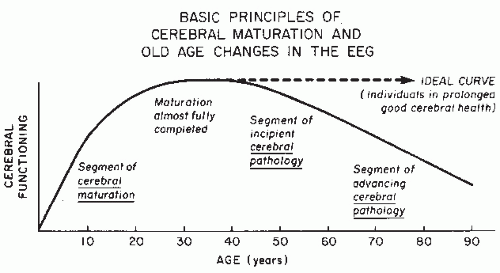
Electroencephalogram (EEG) interpretation depends on accurate pattern recognition. One of the first lessons the novice electroencephalographer learns is that EEG pattern interpretation must take into account the patient age and the level of vigilance, or state. EEG patterns vary according to central nervous system (CNS) development and maturation. This process evolves over time, starting with the early development and maturation of the nervous system (an evolution) to a peak of maturity, followed by an involution. Basic differences exist between the ascending (developmental) and the descending (involutional) portions of this curve. This chapter discusses the influence of age and thus the ontogenetic evolution or maturation of the EEG.

The declining curve of life is not determined by a reverse process such as “dematuration” (certainly not by demyelination), although there is degeneration of CNS structures. CNS aging is caused predominantly by a variety of pathologic processes. The awareness of this basic difference between the ascending and the descending portions of life is of great significance in practical electroencephalography. During the maturational phase, the EEG must be interpreted knowing the conceptual age (CA) and according to statistical norms, whereas in the aged individual, the EEG is interpreted using the ideal or optimal norm, because the optimal situation, even in advanced age, is absence of pathology. Using a statistical norm, a mentally alert octogenarian with the frequency pattern of a normal middle-aged adult would have to be read as abnormal. This would be an absurdity.

Figure 10.1 demonstrates the maturational period, the peak of maturity, and the decline caused by pathology. The peak period of EEG maturation lies well into adulthood (after the age of 30 years), at a time when biologic aging of body tissues has already started and physical capabilities or athletic performances have clearly passed their peak. However, cerebral maturation is not a smooth process of steady growth, since quantum changes occur in prematurity, as well as in infancy and childhood (1).


Figure 10.1 Cerebral maturation (infancy, childhood, and adolescence) versus pathology with regard to repercussions on the EEG.


HISTORICAL ASPECTS

Berger (2) performed the first EEG studies in children and discovered age-dependent changes. The EEG in normal and abnormal children has been well studied and has been expanded with quantified EEG (QEEG). The developmental aspect of EEG interpretation is especially important in the interpretation and study of the neonatal EEG. The technical performance of the EEG and its interpretation in the premature and full-term newborn requires such specialized knowledge that neonatal electroencephalography is now a special branch of electroencephalography, neonatal electroencephalography. Neonatal EEG is described in Chapter 9.

The EEG evolution from infancy to adolescence in a large number of healthy adolescents has been extensively studied by Swedish authors (3, 4, 5, 6 and 7), whose work set the standards for developmental electroencephalography. In a remarkable study of 1416 healthy subjects with an age range from 6 to 39 years using QEEG, Matsuura et al. (8) investigated the development of various frequency ranges over the occipital, central, and frontopolar regions. Gasser et al. (9,10) investigated the development of the EEG in children and adolescents by frequency analysis and topography.

EEG maturation has also been studied in animals. A study in the dog by Pampiglione (11) and the work of Caveness (12) in the rhesus monkey deserve special attention. Table 10.1 provides

a condensed presentation of a number of EEG variables and their developmental aspects.


Table 10.1 A Condensed View of EEG Maturation


Premature (24-27 Wk)


Premature (28-31 Wk)


Premature (32-35 Wk)


Full-Term Newborn (36-41 Wk)


Continuity


Discontinuous, long flat stretches


Discontinuous


Continuous in waking state and REM sleep, discontinuous in non-REM sleep


Continuous except for tracé alternant in non-REM (quiet) sleep


Interhemispheric synchrony


Short bursts in synchrony


Mostly asynchronous activity leads


Partly synchronous, especially in occipital


Minor asynchronies still present


Differentiation of waking and sleeping


Undifferentiated


Undifferentiated


Waking distinguished from sleep early in the period, then differentiation of non-REM and REM sleep


Good


Posterior basic (alpha) rhythm


None


None


None


None


Slow activity (awake)


Very slow bursts, high voltage (state of vigilance undifferentiated)


Very slow activity predominant


Slow (delta) with occipital maximum


Slow (delta), mostly of moderate voltage


Temporal theta


Present and increasing


Prominent


Decreasing and disappearing


Disappearing or absent


Occipital theta


Prominent


Decreasing


Decreasing


Absent


Fast activity (awake)


Very little beta activity


Frequent ripples or brushes around 16 per second


Frequent ripples or brushes (16-20 per second)


Decreasing ripples, sparse fast activity


Low voltage


Long flat stretches


Flat stretches, mainly asynchronous


Low-voltage record suspect of serious cerebral pathology


Very low voltage records are due to severe cerebral pathology; prognosis ominous


Hyperventilation


Not feasible


Not feasible


Not feasible


Not feasible


Intermittent photic stimulation


Unknown


Unknown


Unknown


Driving response below 4 flashes/second may occur, not easily elicited


Drowsiness


Undifferentiated


Undifferentiated


Undifferentiated


Undifferentiated


Tracé alternant


None


None


Present in non-REM (quiet) sleep


Present in non-REM (quiet) sleep


Spindles


None


None (but ripples present)


None (but ripples present)


None (but scanty ripples)


Vertex waves and K complexes


None


None


None


None


Positive occipital sharp transients of sleep


None


None


None


None


Slow and fast activity in sleep


Slow activity of high voltage, little slow activity (stage of vigilance undifferentiated)


Much slow activity, more irregular; little fast activity


Irregular slow activity of occipital predominance


Much delta and theta activity, continuous in REM sleep


REM sleep


Undifferentiated


Undifferentiated


Continuous slow activity; oculographically, REM present


Continuous slow activity, REM in EOG (more REM or “active” than non-REM sleep)


Rhythmical frontal theta activity (6-7 per second)


None


None


None


None


14 and 6 per second positive spikes


None


None


None


None


Psychomotor variant (marginal abnormality)


None


None


None


None


Sharp waves, spikes


Some intermixed sharp activity in bursts (normal)


Some intermixed sharp activity (normal)


Often prominent sharp waves or spikes (normal)


Some minor sharp transients (normal) (abnormal spikes more consistent and prominent)


Infancy (2-12 mo)


Early childhood (12-36 mo)


Preschool age (3-5 yr)


Older children (6-12 yr)


Adolescents (13-20 yr)


Continuous


Continuous


Continuous


Continuous


Continuous


No significant asynchrony


No significant asynchrony


No significant asynchrony


No significant asynchrony


No significant asynchrony


Good


Good


Good


Good


Good


Starting at age 3-4 mo at 4 per second, reaching about 6 per second at 12 mo


Rising from 5-6 per second to 8 per second (seldom 9 per second)


Rising from 6-8 per second to 7-9 per second


Reaching 10 per second at age 10 yr


Averaging 10 per second


Considerable


Considerable


Marked admixture of posterior slow activity (to alpha rhythm)


Varying degree of posterior slow activity mixed with alpha


Posterior slow activity diminishing


None


None


None


None


None


None


None


None


None


None


Very moderate


Mostly moderate


Mostly moderate


Mostly moderate


Moderate, except for low voltage fast records


Uncommon, usually abnormal


Uncommon, usually abnormal


Uncommon, usually abnormal


Seldom as variant of normalcy


Occasionally and (at end of teenage period more often) as variant of normalcy


Not feasible


Mostly not feasible


Often marked delta response


Often marked delta response


Delta responses become less impressive


Improving driving to low flash rates after age 6 mo


Often good driving response to low flash rates


Often good driving response to low flash rates


Often good driving response, chiefly at medium flash rates (8-16 per second)


Often good driving response, chiefly at medium flash rates


Around age 6 mo, appearance of rhythmical theta


Marked “hypnagogic” rhythmical theta (4-6 per second)


Rhythmical theta gradually vanishing, other types of slow activity predominant


Gradual alpha dropout with increasing slow activity


Gradual alpha dropout with low-voltage stretches (mainly slow)


Disappears in first (seldom second) month


None


None


None


None


Appear after second month; 12-15 per second, sharp, shifting


In second year, sharp and shifting, then symmetrical with vertex maximum


Typical vertex maximum


Typical vertex maximum


Typical vertex maximum


Appear mainly at 5 mo, fairly large, blunt


Large, becoming more pointed


Large with an increasingly impressive sharp component


Large with a prominent sharp component


Not quite as large, sharp component not quite as prominent


None


Poorly defined


Poorly defined


Still poorly defined but gradually evolving


Often very well developed


Much diffuse 0.75-3 per second activity with posterior maximum; moderate fast activity


Marked posterior maximum of slow activity; often a good deal of fast activity


Predominant slowing but less prominent posterior maximum


Much diffuse slowing, slightly decreasing voltage


Much diffuse slowing with further attenuation of voltage


REM portion decreasing; mostly slow activity


Mostly slow, starting to become more desynchronized


Slow activity with some desynchronization


Less slowing and increasing desynchronization


Mature desynchronization


None


Seldom in third year of life


May occur, not very common


A bit more common


A bit more common, declining at end of period


None


Rare


May occur, not very common


Fairly common


Fairly common


None


None


Probably none


Uncommon


More common (although relatively rare)


Essential as abnormal phenomena


Spikes in seizure- free children, mainly occipital (mild abnormalities)


Spikes in seizure-free children, mainly occipital, also Rolandic (slight abnormalities)


Spikes in seizure-free children, mainly Rolandic (central-mid-temporal), slight to moderate abnormalities; physiological occipital spikes in congenitally blind children


Benign Rolandic spikes usually disappear before beginning of this period


DISAPPEARANCE OF NEONATAL PATTERNS

Neonatal EEG patterns generally end between 46 and 48 weeks CA and transition to predominantly infantile EEG patterns end by 3 months of age. The trace alternant pattern is the EEG correlate of quiet sleep (non-REM sleep) and is common during the first 1 to 3 weeks of life in a full-term newborn. Active sleep (REM sleep) occurs more commonly in the sleeping newborn (64% of sleeping time, according to Passouant et al. (13)). The healthy full-term newborn demonstrates active sleep (REM sleep) at sleep onset.

Quiet sleep onset is well established 1 month after full-term delivery (37 days, after Ellingson (14)) and gradually emerges as the predominant type of sleep with the development of stages 2, 3, and 4. Trace alternant patterns disappear about 3 to 4 weeks after full-term birth (15), although they have been observed up to day 47 (14). Intervening CNS insults, either primary or secondary (systemic), may interfere with CNS function and temporarily cause developmental regression, which may be expressed with a return to a less mature EEG pattern.

With the disappearance of neonatal EEG patterns and the emergence of patterns such as the posterior basic (the posterior dominant rhythm), the forerunner of alpha rhythm, and sleep spindles, the EEG begins to demonstrate the more mature patterns seen in adults. During the waking state, a generally rhythmical 3 to 4 Hz posterior activity occurs, a precursor of the posterior alpha rhythm, or the posterior occipital or dominant rhythm. This posterior rhythm demonstrates reactivity to eye opening and closing: blocking with eye opening and activation with eye closing.


EEG IN INFANCY (2 TO 12 MONTHS)


General Considerations and Technical Aspects

It is not easy to obtain a waking EEG recording in an infant. As infants tend to keep their eyes open, in order to obtain a posterior dominant rhythm, Dreyfus and Curzi-Dascalova (15) suggest gentle passive occlusion of the eyes (passive eye closure) for short periods to activate the posterior basic rhythm. Temporary use of a short-time constant (0.1 second) may be helpful to separate the posterior rhythm from movement artifacts. This rhythm may also be present in a crying infant (associated with forceful closure of eyes and concomitant frontal muscle artifacts), as well as in a quiet infant with open eyes. The infant usually closes the eyes as a sign of impending drowsiness.

There are two basic philosophies to EEG recording in infancy. In the first, the recording is started in a sleeping baby. Spontaneous sleep is desirable, and this may be achieved when the recording is scheduled shortly after feeding. In the past, sedation was commonly used, usually chloral hydrate. Because of concerns for adverse events with conscious sedation, such as apnea, the use of conscious sedation for EEG has been disappearing. Many laboratories now favor sleep deprivation the night before and allow the infant to lie on a bed or in the mother’s arms. Sweeney et al. (16) compared sleep deprivation versus sedation. The recordings had fewer artifacts and were off a higher quality with sleep deprivation. Guidelines have been established for sleep deprivation (17). The use of sleep deprivation has been confirmed by Ong et al. (18). In a sedated infant, it is often impossible to secure a waking tracing. Using the second approach, the record routinely starts while the baby is awake. It is extremely difficult to place electrodes under these circumstances as most infants resent this type of manipulation.

Some EEG technologists have become extremely experienced in the art of handling a waking infant, and often succeed in obtaining both waking and spontaneous sleep records. Further information about the management of infants and children in North American EEG laboratories is found in the study of Leonberg (19).

The International 10-20 Electrode System should be used for electrode placement, using a full set of 21 EEG electrodes, 19 scalp electrodes, and two ear reference electrodes, according to the American Clinical Neurophysiology Society guidelines (20). The use of ocular leads, respiratory monitoring, and electrocardiogram (ECG) are helpful in the detection of artifacts, as well as in the assessment of sleep stages.

Although some laboratories use rubber caps in small infant sizes, this technique is not conducive to sleep, and collodion is preferable, especially in the larger pediatric hospitals. Bentonite paste (recommended by Fois (21)) is no longer available. Excellent new types of commercial paste are now available, but when used, it is possible that rapid drying and thus deteriorating electrode function may occur, resulting in high electrode impedance values, in addition to the natural higher impedance of the scalp in newborns and early infancy. Needle electrodes are now rarely used.


EEG Characteristics in the Waking State

In early infancy (around the age of 2 months), irregular delta activity of 2 to 3.5 Hz and medium to high voltage (50 to 100 µV) is widely preponderant. As noted above, rhythmical occipital 3- to 4-Hz activity is often noted at the age of 3 to 4 months; this activity can be blocked by eye opening.

This posterior basic (dominant) rhythm becomes more stable at the age of 5 months and increases over time, first to 5 Hz (15) and subsequently to an average frequency of 6 to 7 Hz (occasionally 8 Hz) by age 12 months. The amplitudes range from 50 to 100 µV.

Some rhythmical rolandic (central) activity of 5 to 8 Hz may be present as early as the age of 3 months, with an amplitude around 25 to 50 µV. This activity is the precursor of the mu rhythm and is stable during the first year of life (15), but may be dependent on somatosensory stimulation (22). These visual analysis findings have been confirmed with QEEG (23, 24 and 25).


EEG Characteristics in Drowsiness

Prior to the age of 5 to 8 months, the transition from the waking state to sleep is a gradual process characterized by progressive slowing into the delta frequency range. No specific drowsy state is identified in this smooth progression of slow activity to the sleep stage. Drowsiness is recognized by a distinct pattern
between the age of 6 and 8 months: a hypersynchronous rhythm in the lower theta range, around 4 Hz, with gradual acceleration to 5 and 6 Hz over the ensuing months. This impressive theta rhythmicity is known as “hypnagogic hypersynchrony” (26).

This drowsy pattern seems to develop from the posterior basic (dominant) rhythm. The transition from wakefulness to drowsiness is associated with a change in the amplitude distribution. The maximum rhythmical theta activity moves into the centroparietal region where amplitudes commonly reach 100 to 250 µV. EEG amplitude values depend on the interelectrode distance and may vary from montage to montage and are best measured with a reference montage.

According to Dreyfus and Curzi-Dascalova (15), the occipital rhythm may be somewhat slower than the diffusely predominant theta rhythm (possibly indicating a basic difference between two coexisting rhythmical theta patterns). In rare cases, the hypnogogic hypersynchrony may not occur.


Sleep EEG and Non-REM Sleep

During the first 3 months of life, sleep may begin in a peculiar manner. Curzi-Dascalova et al. (27) have shown that some infants fall asleep without eye closure, others with half-closed eyes, and others with brief alternating opening and closing of the eyes. The EEG and polygraphic data indicate sleep onset with active (REM) sleep in neonates and gradual evolution of sleep onset with quiet (non-REM) sleep during the ensuing weeks. The “slow” sleep of the infant is dominated by diffuse 0.75- to 3-Hz activity with a maximum amplitude (100 to 150 or 200 µV) over the occipital area, “occipital delta.” This occipital delta activity may be quite prominent during the first year of life. The amplitudes increase with deepening slow sleep. There are some intermixed theta, alpha, and beta frequencies of smaller amplitudes.

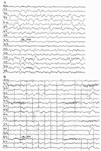
Sleep spindles usually appear during the second month of life; occasionally, spindle fragments may be seen somewhat earlier (28). The spindle frequency ranges from 12 to 15 Hz, with 14 Hz the most commonly encountered frequency. Throughout infancy, spindles are maximal over central and parietal areas with shifting asymmetries. A clear-cut midline (vertex) maximum does not exist at this age. Spindles of infancy usually show a negative sharp component, whereas the positive component is rounded. The sharp spindle configuration (shown by Fois (21)) is a typical hallmark of sleep in infancy. The comb-like shape of these runs may be erroneously interpreted as 14-Hz positive spikes, but a careful analysis of polarity clearly shows the negativity of the spiky components. The spatial distribution also is different: spindles are rolandic, whereas 14-Hz positive spikes are predominant in a posterotemporal location. Finally, the 14-Hz or 14- and 6-Hz positive spike pattern is extremely rare before the age of 2 years and virtually nonexistent during the first year of life (Fig. 10.2).

Spindles may show sharp negative and positive phases after the age of 6 months Katsurada (29). This sharp positive spindle polarity is less common than spindles with a strictly negative sharp component.

The spindle train duration varies with age. Spindle trains are of short duration and low voltage during the second month. With the third month, the amplitude increases and the duration of the runs becomes much longer (30). The intervals between spindle trains become shorter, but we do not agree that spindling is almost continuous between the age of 2 and 6 months (24). Spindle bursts may reach a duration of 10 seconds in the second half of the first year, but there is a decreased duration of each spindle run, while the overall number of runs increases.

Dreyfus and Curzi-Dascalova (15) report that the complete absence of spindles at the age of 3 to 8 months represents a severe abnormality. However, this finding should be interpreted with great caution, as enough sleep must have been obtained during the recording in order “to give the baby a chance to produce spindles.” At this age, there is still a fair chance that the sleep recording is limited to REM sleep; this demonstrates the importance of oculographic and pneumographic recording.

An excellent demonstration of the development of sleep spindles from the age of 10 weeks to 1 year was presented by Hughes (31) (Fig. 10.3).

Vertex waves and K complexes are usually seen around the age of 5 months, although rudiments may occur much earlier. Vertex waves may be quite large at the age of 5 to 6 months. At this age, K complexes are of considerable voltage, but the initiating sharp component is poorly developed and somewhat “blunted” (32). In contrast, Metcalf et al. (33) have stressed the comparatively low voltage of K complexes in infancy. These authors also noticed the appearance of K complexes in infants aged 5 to 6 months, but the complexes may be obscured by background activity (Figs. 10.4 and 10.5). A vertex wave may be frontally dominant or extend into the lateral frontal regions; this is called an F-wave (34) (Fig. 10.6).

In normal infants in the first year of life, Ellingson et al. (35) recorded brief apneic episodes (“respiratory pauses” lasting 3 to 10 seconds) during sleep. These pauses are more common in REM sleep.


Sleep EEG and REM Sleep

REM sleep abundance decreases during the first year of life (36,37), evolving from approximately 50% at birth, falling to 40% at 3 to 5 months and 30% between 12 and 24 months.

Dittrichova et al. (38) and Dreyfus and Curzi-Dascalova (15) reported the occurrence of sharply contoured occipital activity in the REM sleep of infants. This activity shows a frequency around 2 Hz at 6 weeks and 2 to 4 Hz at 12 to 16 weeks of age. There is some intermixed delta and theta activity associated with the occipital sharp transients.

The REM sleep latency (time span from sleep onset to the first REM period) gradually lengthens during the first year of life. Schulz et al. (39) has shown that the REM sleep latency underlies diurnal rhythmical changes, with the longest latencies between noon and 4 PM and the shortest between 4 AM and 8 AM.


Reactivity and Evoked Responses

The evolution of evoked responses in newborns and infants are discussed in Chapter 49.

The blocking response of the posterior basic (dominant) rhythm (obtained by passive eye closure, see General Considerations and Technical Aspects)
is usually present at the age of 3 to 4 months. The central mu rhythm does not react to eye opening, which permits differentiation when there is anterior spreading of the posterior dominant rhythm. A true rolandic mu rhythm cannot be identified during the first year of life, although forerunners of this activity are likely to be present. The authors’ earliest observation of unmistakable rolandic mu rhythm was made in a 20-month-old child (40).


Figure 10.2 A: Patient age 10 months. Light non-REM sleep. Typical spindles of infancy, sharp and shifting. Normal posterior voltage maximum of slow activity (channels 3 to 7 from the top). B: Patient age 9 months. More prolonged trains of infantile spindles.

A photic driving response to flickering light may be obtained as early as 3 to 4 months after delivery; the responses are most prominent in the theta band (41,42). Occipital lambda wave activity occurs rarely during the first year of life.

Only gold members can continue reading. Log In or Register to continue

Stay updated, free articles. Join our Telegram channel

Sep 9, 2016 | Posted by in NEUROSURGERY | Comments Off on Normal EEG and Sleep: Infants to Adolescents

Full access? Get Clinical Tree

Get Clinical Tree app for offline access