Fig. 13.1
(a, b) A 15-year-old male with DMD and delayed presentation of his progressive scoliosis. His lumbar curve is 128° and he has significant pelvic obliquity. (c, d) The patient underwent posterior spinal instrumentation and fusion from T3 to the pelvis with iliac screws. To assist with correction, intraoperative traction as well as multilevel Ponte type osteotomies were performed
There are similar choices for instrumentation to the pelvis. Options include the Galveston techniques with either Luque or Unit Rods, Dunn-McCarthy technique with an S-rod, sacral screw, and iliac screw fixation [42, 44, 53–55]. Each has advantages and disadvantages. Galveston technique is subject to loosening and migration of the rod [11]. In addition, the Galveston technique sometimes requires complex three-dimensional contouring to fit the altered pelvic anatomy. Iliac screws, on the hand, are placed individually into each iliac wing and then connected to the rod through connectors. A recent study by Peelle et al. demonstrated equal effectiveness in controlling pelvic obliquity between the Galveston technique and iliac screw fixation [55]. Our preferred method is to utilize iliac screws when instrumenting to the pelvis in DMD patients.
Another important consideration in the preoperative planning for scoliosis is the risk of blood loss during surgery. Of all pediatric spine surgeries, Duchenne muscular dystrophy has demonstrated, on average, to have the highest mean level of blood loss [56, 57]. This is important considering their poor cardiac reserve. These patients require a large exposure from the upper thoracic spine to the lower lumbar spine or pelvis. The paraspinal muscles are difficult to elevate subperiosteally. Dysfunction of vascular smooth muscle as well as decreased platelet adhesion is thought to contribute to increased blood loss [57, 58]. Besides diligent hemostasis intraoperatively, the use of antifibrinolytics may help to minimize the blood loss. Shapiro et al. retrospectively evaluated the use of transexamic acid in 20 DMD patients and compared them with 36 control patients [59]. Transexamic acid was found to reduce intraoperative blood loss and the need for homologous transfusions. Other options which have been published for adolescent idiopathic scoliosis but not DMD include the use aminocaproic acid [60–62]. Vitale et al. investigated the efficacy of preoperative erythropoietin on hematocrit and transfusion rates in neuromuscular patients. They found no clinical benefit in their treatment group. We currently work with anesthesia preoperatively to ensure that each patient is administered an antifibrinolytic during surgery. Intraoperative blood loss is also collected in a cell saver and given back to the patient. Postoperatively, hematocrits are monitored closely to ensure that cardiac function is not overly stressed.
13.1.5 Long-Term Outcomes
As previously discussed, there is controversy whether scoliosis surgery improves pulmonary function in the DMD patient. A recent Cochrane Review by Cheuk et al. was unable to provide an evidence-based recommendation for scoliosis surgery in DMD. Their reasoning was the lack of randomized clinical trials. Of the 36 relevant studies addressing the outcomes of scoliosis surgery, none met the inclusion criteria for review.
Studies have suggested that scoliosis surgery does benefit patients beyond pulmonary function [63–65]. Bridwell et al. sent questionnaires to 33 patients with DMD evaluating function, self-image, cosmesis, pain, quality of life, and satisfaction [63]. Patients reported benefits in all categories with the highest ratings in cosmesis, quality of life, and satisfaction. Granata et al. and Takaso et al. found that sitting position, aesthetic improvement, and quality of life were all improved following spinal fusion [52, 64]. More than 90 % of their patients/parents would give their consent again for surgery.
13.1.6 Summary
Spinal deformity commonly affects the male patient with DMD. Treatment of this deformity is complicated by the progressive muscle weakness and deteriorating pulmonary function. Current literature suggests that surgical management of the deformity can maintain upright sitting posture, improve quality of life, and positively affect short-term pulmonary function. Unfortunately, a lack of randomized controlled trials has prevented any formal evidence-based recommendation from being made by a Cochrane Review. If surgery is contemplated, however, it should be performed early when the patient is at his or her maximal health. In addition, if there is more than mild pelvic obliquity, one should consider including the pelvis in the instrumentation and fusion.
13.2 Spinal Muscular Atrophy
Initially described by Guido Werdnig, spinal muscular atrophy (SMA) is a rare autosomal recessive disorder characterized by degeneration of the anterior horn cells of the spinal cord and the neurons of the lower bulbar nuclei [66, 67]. Two genes are associated with this disease: the survival motor neuron gene and the neuronal apoptosis inhibitory protein gene [68]. SMN protein appears to interact with RNA-binding proteins and is found in both the nucleus and cytoplasm of cells [69]. It is considered the most common fatal neuromuscular disease of infancy and the most common neuromuscular disease in children [66].
13.2.1 Classification
Common to all SMA patients is a symmetric muscular weakness predominantly affecting the lower limbs and proximal muscles compared with the upper limbs or distal muscles. Patients usually have normal intelligence with no effect on sensibility. The age of onset and clinical course can have a variable presentation. Due to this heterogeneity, spinal muscular atrophy is most commonly divided into three types [70–72].
13.2.1.1 Type I, Acute Werdnig-Hoffman Disease
Type I SMA is the most severe form of the disease, usually presenting at birth or within the first 2–6 months of life. These patients do not meet early motor milestones with the inability to gain head control, roll over, or sit up. It has been suggested that in utero osteoporosis from decreased movement is responsible for post-natal pathologic fractures [73]. Patients with Type I SMA usually do not survive beyond the age of 3 years. Respiratory failure from intercostal weakness and rib collapse is responsible for their mortality. Due to their early mortality, orthopedic intervention is rarely indicated in these children.
13.2.1.2 Type II, Chronic Werdnig-Hoffman Disease
The clinical onset of Type II SMA occurs between the ages of 6 and 24 months. Patients reach early motor milestones but are never able to walk independently. Weakness usually starts in the lower extremities, affecting the gluteal and quadricep muscles initially. Life expectancy is variable from adolescence to adulthood with some patients living into their fourth decade [66, 74]. The cause of mortality is respiratory failure.
13.2.1.3 Type III, Kugelberg-Welander Disease
The clinical onset of Type III SMA occurs following the age of 18 to 24 months. In nearly all cases, the diagnosis is made before the age of 10 years. As expected, children attain greater motor milestones compared with Type II SMA. Patients are able to walk independently until early adolescence. Russman et al. reported that 50 % of those children with an age of onset before 2 years lost their ability to walk without assistance by age 12 [75]. Those children that presented after age 2 typically were ambulating into the fourth decade. Patients that never reached independent ambulation lost their ability to walk by age 7.
13.2.1.4 Functional Classification
Evans et al. described a functional classification based on the maximum physical function achieved [76]. The purpose was to give insight into the patient’s prognosis. Group I patients never sit independently, have poor head control, and develop early progressive scoliosis. Group II children have head control and ability to sit but cannot walk or stand. Group III patients can stand by themselves and are able to walk with external support. Group IV children can walk and run independently.
13.2.2 Diagnosis
For those patients that do not present at birth, presenting concerns by families are a delay in reaching motor milestones. Depending on the age of the patient, these include an inability to gain head control, roll over, sit, stand, or walk independently. Physical examination should then assess motor strength as well as deep tendon reflexes. For those patients that present early (Type I or II), gross fasciculations of the tongue or tremors of the finger are commonly present [66, 77].
Once SMA is suspected, further diagnostic workup includes laboratory studies, nerve conduction studies, electromyography (EMG), and DNA testing. Creatine phosphokinase and adolase are usually normal or slightly elevated in Type III patients [78]. Motor and sensory nerve conduction velocities are normal. EMG findings demonstrate fibrillation potentials associated with denervation as well as large polyphasic motor units associated with renervation [66, 78]. DNA testing is highly sensitive for SMA with PCR the diagnostic procedure of choice [79]. Muscle biopsy is also highly diagnostic. Histologic findings include muscle fiber degeneration and atrophy with no evidence of primary myopathy [78].
13.2.3 Spinal Deformity
Scoliosis is the most common orthopedic problem in patients with SMA [80]. Nearly 100 % of Type II SMA patients and half of Type III SMA patients develop a spinal deformity [76, 81–83]. The deformity is typically a right-sided C-shaped thoracolumbar curve. Ninety percent of the patients have a single curve. The curve is usual progressive and in approximately a third of the cases associated with a progressive kyphosis [81, 82]. Similarly to DMD patients, development of scoliosis in type III SMA occurs with their loss of ambulation [80]. Pulmonary function is similarly compromised in patients with SMA [84]. The worsening of lung function is secondary to muscle weakness as well as the progressive scoliosis.
As there is a difference in the clinical presentation between the three types of SMA, there is similar heterogeneity in the risk and progression of scoliosis. Evans et al. demonstrated that the age of scoliosis onset correlated with the severity of muscle disease [76]. Type I SMA patients typically had scoliosis by the age of 2 years, while Type III SMA patients developed scoliosis between the ages of 4 and 14 years. The rate of progression was also highly associated with the disease severity ranging from 8.3° per year in severe cases to 2.9° in more mild cases.
As for the severity of the scoliosis, a study by Granata et al. reported curves ranging from 10 to 165° [81]. Schwentker and Gibson reported on 50 patients with SMA [80]. Seventy percent had scoliosis measuring greater that 20°, and 40 % had curves greater than 60°. The natural history of these large curves suggests that they can be quite disabling [76]. In addition to trouble sitting, patients can lose upper extremity function to maintain trunk balance as well as develop back pain or pain from rib impingement on the pelvis.
13.2.4 Nonsurgical Management of Scoliosis
Orthotics has generally been thought to be ineffective in preventing the development or progression of neuromuscular scoliosis [81, 85, 86]. It, however, has been shown to be effective in improving sitting balance. Letts et al. demonstrated an improvement in sitting stability in 80 % of patients with a collapsing neuromuscular scoliosis with the use of a soft Boston orthosis [87]. They also thought that a soft brace was more tolerable than a rigid orthosis and resulted in less skin breakdown.
Some studies have suggested that the use of orthotics may slow the rate of progression of scoliosis [82, 86, 88]. Slowing the rate of scoliosis progression has the advantage of allowing patients to get older when they are more suitable for a surgical intervention. This is especially critical in the early-onset patients (Type I and II SMA). Unfortunately, most of these studies report opinion and have not given reliable data to demonstrate that bracing truly slows the progression of spinal deformity. Bracing is also not without its morbidity. Aprin et al. reported on five patients that had to discontinue their brace secondary to respiratory difficulty [86].
Our preferred nonsurgical treatment is to initiate bracing in patients with spinal deformities on sitting films between 30 and 40°. Typically, the curves in SMA are quite flexible and amenable to the orthosis. We find that the brace in addition to wheelchair supports help to maintain sitting balance. This is especially critical in pre-adolescent patients where attempts are made to delay surgery until the patient is more mature. In some cases, especially in the Type I patient where long-term survival or surgical tolerance is not expected, bracing may be the definitive management of the spinal deformity.
13.2.5 Surgical Management of Scoliosis
Similarly to other neuromuscular scoliosis, the decision to operate on an SMA patient is dependent on multiple factors. In general, the radiographic parameters for spinal fusion are not controversial and simple to follow. We recommend spinal fusion for curve magnitudes greater than 50° that are refractory to conservative measures and demonstrate progression. These indications for surgical fusion have been recommended by other authors as well [66, 76, 89].
Unfortunately, patient factors may not make the above rules simple to follow. In some patients with type I SMA, their early-onset scoliosis and grim long-term survival have made surgical intervention unreasonable. Type II patients may also present with a progressive scoliosis at an early age. Spinal arthrodesis would have a considerable negative effect on trunk growth as well as lung growth. These patients are also at significant risk of developing a crankshaft deformity necessitating an anterior fusion [90]. In these cases, spinal fusion is indicated, but an attempt at delaying surgery with the use of an orthosis is made. The goal is to maintain some control of the curve until a definitive procedure can be done at about the age of 10. Of course, this may mean watching a curve progress to greater than 80°.
13.2.5.1 Growing Spine Techniques
Previously, there has been some thought about using an expandable or “growing rod” construct in young SMA patients that developed significant scoliosis. Fujik et al. reported using an expandable or “telescoping” device in type II SMA patients [91]. The device was abandoned due to its technical demands and inability to prevent progression of the deformity and crankshafting. They concluded that a brace should be used until the age of 10 when a fusion can be performed.
However, more recent publications on early-onset scoliosis in SMA suggest increasing use of growing spine constructs. Growing rods may be the answer for these young patients. Chandran et al. found excellent deformity correction in 11 patients (mean age of 6 years) from 51° to 21.6° with a low complication rate at the initial surgery [92]. Sponseller et al. demonstrated in six patients that growing rods can be fixed to the pelvis and result in improved coronal balance, sagittal balance, and pelvic obliquity [93]. With similar constructs, McElroy et al. found improved major curve by nearly 50 %, improved trunk height, and improved space available for lung ratio at final follow-up. However, they did not find any halt in rib collapse, which is common in SMA. Additionally, they found that patients with SMA had longer hospital stays than did patients with early-onset idiopathic scoliosis undergoing the same procedure [94]. Recently, Tobert and Vitale published a case series of three SMA patients (aged 8, 7, and 3 years) undergoing rib to pelvis growing construct. They found stabilization of pulmonary function and overall improvement in quality of life and caregiver burden [95].
Additional literature is needed to know the long-term benefits and complications related to these surgeries. The tolerance for multiple anesthetics needed to expand the device on these already pulmonary compromised patients is also not known. To minimize this need for multiple anesthetics, we have utilized a modified Shilla technique to manage the early severe scoliosis (Fig. 13.2).
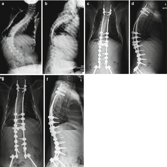
Fig. 13.2
(a, b) An 8-year-old female with SMA and progressive kyphoscoliosis. Her coronal major curve measures 90°. (c, d) The patient underwent a modified Shilla technique with instrumentation and fusion from T10 to the pelvis. The instrumentation was extended proximally to pedicle screws at T3 and T4 with fusion across these two levels. The pedicle screws are allowed to slide along the rod as the spine grows. The goal of this procedure is to attain correction with the distal fusion but allow thoracic growth with the Shilla technique. (e, f) At 1 year postoperative, the patient had grown nearly 1 cm as measured by the movement of the top screws
13.2.5.2 Posterior Spinal Fusion
In the older SMA patient that requires definitive spinal stabilization, the standard is PSF and SSI. The goal is to prevent progression and obtain an alignment that will improve or maintain balance and sitting ability. In the non-ambulatory patient, this typically involves segmental instrumentation from T2 to the pelvis. Many spinal deformity surgeons report good outcomes using sublaminar wires with Luque rods or a unit rod for the treatment of neuromuscular scoliosis [40, 83, 96]. Others are transitioning to the use of pedicle screws to provide more rigid fixation [40]. The improved fixation to bone with pedicle screws has decreased the use of postoperative bracing for some neuromuscular patients [66]. We continue to brace all neuromuscular patients for 3 months postoperatively to prevent excessive stress on the osteopenic bone during transfers, including those with all pedicle screw instrumentation. Pelvic instrumentation is recommended to prevent progressive pelvic obliquity and difficulty with sitting [80]. Similarly to patients with DMD, options include Galveston technique or iliac screws.
The use of an anterior approach has traditionally been reserved for severe curves or for patients at risk of developing crankshaft deformity. In the case of patients with SMA, other factors need to be considered. These patients typically have poor pulmonary reserve associated with weakness of their respiratory muscles. This places them at increased risk of developing pulmonary complications. The use of segmental fixation may decrease the risk of crankshaft deformity. Smucker and Miller reported on 43 patients with neuromuscular scoliosis and open triradiate cartilage treated with a unit rod [97]. They found no evidence of crankshaft deformity at 2-year follow-up. Some believe that pedicle screws may further decrease the risk by providing three-column fixation. However, this needs to be evaluated. There is also increasing evidence that severe spinal deformity can be completely managed from a posterior approach. Multilevel posterior osteotomies or single-level vertebral column resections stabilized with pedicle screws have been shown to adequately treat the severely deformed, rigid spine [98]. However, this, too, has not been adequately studied in patients with spinal muscular atrophy.
In preparation for spinal fusion, all patients with SMA should be evaluated by a pulmonologist, neurologist, and anesthesiologist. This will ensure that patients are optimized for surgery especially regarding their pulmonary function. In the immediate postoperative period, patients are most at risk of developing pulmonary complications. Aprin et al. reported a 45 % incidence of respiratory problems following surgery [86]. Four of their 22 patients required intubation. Brown et al. reported that tracheostomy was needed in 30 % of their patients [96]. The use of preoperative traction has been suggested to increase spinal flexibility and improve pulmonary function, possibly diminishing their risk of respiratory complications [86, 99]. Postoperatively, these patients should have aggressive pulmonary therapy and early mobilization. Ventilatory assistance with the guidance of a pulmonologist may be needed several days following the surgery. Other long-term complications following spinal arthrodesis include crankshafting, pseudoarthrosis, prominent implants, narrowing of the chest, gastric volvulus, and diaphragmatic rupture [63, 82, 86, 100]. Except for crankshafting, these complications were more commonly seen in older patients with larger deformities.
13.2.6 Long-Term Outcomes
In general, the literature supports spinal fusion in SMA patients with progressive scoliosis. Multiple authors have reported improvements in sitting, balance, comfort, and cosmesis [81, 99]. Bridwell et al. evaluated 21 SMA patients with an average follow-up of 7.8 years after surgery [63]. Patients reported benefits in all categories with the highest ratings in cosmesis, quality of life, and satisfaction. In contrast, some authors have reported a decline in some functional activities, specifically upper extremity activities. Brown et al. demonstrated a decline in self-feeding, drinking, and self-hygiene at 2-year follow-up with some improvement at 5 years [96]. Furumasu et al. reported similar findings suggesting that the lack of spinal flexibility diminished gross upper extremity motor function due to a change in trunk position. What is unclear in these patients is the influence of a progressive muscle disease in the diminished functional activities.
Pulmonary function also appears to benefit from stabilization of the scoliosis. Robinson et al. demonstrated a significant improvement in lung function in the patients that underwent spinal fusion [84]. They also demonstrated a significant inverse linear relationship between curve magnitude and percentage of predicted vital capacity.
13.2.7 Gene Therapy
Spinal muscle atrophy is caused by a mutation in the survival motor neuron (SMN) 1 gene that results in a reduction of the SMN protein. Patients also can have variations in the copies of the SMN2 gene, which produces reduced levels of SMN protein. This production, however, is insufficient for normal motor neuron function [101]. There are promising gene therapy pathways that are being tested to increase the number of SMN proteins produced [102, 103]. One approach is to antisense oligonucleotides to redirect SMN2 translation and increase production of fully functional SMN protein [104].
13.2.8 Summary
Spinal muscle atrophy is a heterogeneous disease commonly affected by progressive scoliosis. Depending on the severity of the disease, patients can have significant deformity at a very early age. While ineffective at preventing scoliosis, bracing is utilized to delay surgery. When severe scoliosis develops at a young age (<10 years), growing spine constructs can improve spinal deformity and sitting balance and may improve pulmonary function and quality of life, but this needs further study. The gold standard for spinal stabilization remains posterior spinal instrumentation and fusion. Current literature suggests that surgical management of the deformity can maintain upright sitting posture, improve quality of life, and positively affect pulmonary function. Whether this improvement in pulmonary status improves life expectancy is still unclear.
13.3 Arthrogryposis Multiplex Congenita
Arthrogryposis or “arthrogryposis multiplex congenita” (AMC) is a heterogeneous group of diseases with the similar phenotype of multiple congenital joint contractures [105, 106]. Currently, there are more than 150 subtypes that result from a failure of normal movement in utero. The etiology for this lack of movement may be myopathic, neurologic, or secondary to connective tissue abnormalities [107]. Amyoplasia is the term used to describe the more classic disease entity seen in orthopedics. These patients have a dysgenesis of anterior horn cells resulting in replacement of muscle with adipose and fibrous tissue [108].
Patients with arthrogryposis multiplex congenita (AMC) have significant musculoskeletal deformities secondary to the contractures. The majority of patients have all four limbs involved (84 %) [105]. Severe equinovarus feet, hip dislocations (unilateral or bilateral), and scoliosis are commonly seen. Non-orthopedic abnormalities include hypoplasia of the labial folds, inguinal hernias, abdominal wall defects, cryptorchidism, gastroschisis, and bowel atresia [105].
13.3.1 Spinal Deformity
The incidence of scoliosis in AMC is reportedly between 30 and 67 % depending on the definition used [109, 110]. The deformities are similar to other neuromuscular conditions with lumbar and thoracolumbar curves predominating [111, 112]. The curves are frequently stiff. Progression of the deformity can be rapid, up to 6.5° per year [112]. The earlier the presentation of scoliosis, the more severe the curve may become and be associated with pelvic obliquity. Increased lordosis is frequently seen.
Scoliosis is typically refractory to orthotic management [111, 112]. Patients with arthrogryposis will frequently develop scoliosis early in life. Little literature has evaluated the treatment of early-onset scoliosis in these patients. Recently, Astur et al. and the Chest Wall Spinal Deformity Study Group evaluated ten children with arthrogryposis that underwent treatment with the use of the vertical expandable prosthetic titanium rib (VEPTR) device and found it to be an effective treatment method in these patients [113]. Using this rib-based distraction device, they obtained 37 % correction of scoliosis and 29 % correction of kyphosis. They also found improved thoracic volume. Six complications occurred in four patients in a total of 62 procedures performed. Proximal junctional kyphosis appeared to remain a problem, however, in this cohort. Other than this series, few studies have evaluated growing spine techniques in patients with AMC.
PSF and SSI remains the standard and appears to be effective in preventing progression of the scoliosis. However, correction of the curves appears to be modest, about 35 % [111]. Yingsakmongkol and Kumar reported slightly increased correction (44 %) with a combined anterior and posterior fusion [109]. These series are dated, however, and do not assess surgical outcomes with current segmental instrumentation. In some cases, instrumentation was not used. If pelvic obliquity is present, fusion to the pelvis should be attempted. Care should also be taken when positioning patients. Their stiff joints and osteopenia place them at increased risk of developing pathologic fracture.
Related posts:
 Repetitive Anesthesia Concerns in Early-Onset Scoliosis
Treatment of Spinal Deformity in Cerebral Palsy
Convex Growth Arrest for Congenital Scoliosis
Management of Spine Tumors in Young Children
Growth Modulation Techniques: Titanium Clip-Screw Implant System (HemiBridge)
Single and Dual Traditional Growing Rods
Repetitive Anesthesia Concerns in Early-Onset Scoliosis
Treatment of Spinal Deformity in Cerebral Palsy
Convex Growth Arrest for Congenital Scoliosis
Management of Spine Tumors in Young Children
Growth Modulation Techniques: Titanium Clip-Screw Implant System (HemiBridge)
Single and Dual Traditional Growing Rods
Stay updated, free articles. Join our Telegram channel

Full access? Get Clinical Tree


