Fig. 40.1
(a) Thoracolumbar scoliosis and (b) concurrent sagittal plane deformity in a 4-year-old male patient who was treated with uninstrumented anterior convex growth arrest. Four-year follow-up radiographs show almost complete correction of (c) coronal spine deformity with (d) slight improvement of the sagittal deformity
As can be learned from the above discussion, none of the variables including the age, type of the anomaly, presence of sagittal deformity or intraspinal anomaly, and length of the curve were found to significantly affect the outcome of the procedure.
40.5 Problems
CGA is accepted as a safe procedure that generally does not result in serious complications except for minor infections (wound or chest) and traction neuropraxias of either the intercostal or cutaneous thigh nerves which are related to anterior surgery [2, 4, 11, 16, 17, 29, 32, 34, 38, 39]. More significant problems include unpredictability of the curve behavior after the procedure and incapability to control the spinal balance.
40.6 Proposed Solutions and Modifications
For each of the drawbacks mentioned previously, potential solutions were proposed by a number of authors. Bandi et al. [3] reported a modified technique on two patients, which spares the segmental vessels during the anterior epiphysiodesis surgery in order to decrease the neurological complications. It has been argued that as the number of segmental vessel ligation increases, there is a corresponding increase in the risk of spinal cord ischemia and there are reports of such spinal cord injuries. In the proposed technique, segmental vessels are mobilized and elevated during the anterior epiphysiodesis procedure. This method provides a method of sparing segmental arteries however it does not address the other potential problems with anterior surgery and thoracotomy.
Keller et al. [16] and King et al. [18] reported an alternative method which avoids the anterior surgery and thus the risks related to it. This method consists of a posterior approach utilizing transpedicular curettage of the end plates anteriorly, from a posterior approach. Posterior hemiepiphysiodesis is done as in the original technique. Transpedicular approach has potential advantages over the standard two-staged operation in that it decreases the neurovascular complications by avoiding the anterior approach. On the other hand, a potential disadvantage is the chance for an incomplete hemiepiphysiodesis of the anterior end plates.
More recently transpedicular approach with short segment posterior instrumentation was reported by Ginsburg et al. [13]. Their series consisted of ten patients with a mean follow-up time of 29.7 months. They reported either no improvement or a decrease in the curves in seven of their patients. Conclusions were drawn that CGA with a transpedicular approach is an effective method for congenital scoliosis especially when done earlier in premenarchal patients and patients with open triradiate cartilages.
Cheung et al. [6] added a growing rod to the concave side and reported their results using posterior convex hemiepiphysiodesis with concave distraction. Distractions were not done in a regular basis but were done only when loss of distraction force was evident. The authors stated that the loss of distraction was manifested by the increasing space between the hook and the C-ring of the Harrington rod or between the hook and the lamina or when the hook became dislocated or the curve deteriorated. At a mean follow-up of 10 years, the authors reported 41 % correction of scoliosis. The concave distraction produced immediate improvement in the coronal balance, and further correction with consecutive distractions was minimal. They suggested that this procedure could be recommended for children with severe deformities and decompensation in the lower thoracic spine.
Another modification that has been proposed by the Hacettepe group is the addition of posterior instrumentation with transpedicular screws to the convex hemiepiphysiodesis (Fig. 40.2a–f) [8]. Specifically, the technique exploits the concept that pedicle screws may control the growth of the vertebral column in both longitudinal [19] and transverse planes [7] as demonstrated in animal studies. Transpedicular screws were placed in all anomalous vertebrae to be hemiepiphysiodesed to eliminate the need for an anterior surgery. Compression-rotation maneuvers were employed to correct the deformity. Added posterior instrumentation provided an initial correction, thereby decreasing the unpredictability of the outcome; however, trunk balance was not achieved in every case.
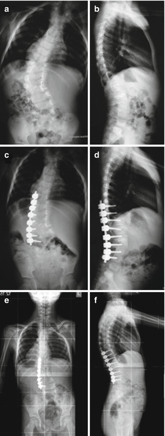
Fig. 40.2
A six-year-old male with (a) thoracolumbar coronal deformity and (b) normal sagittal alignment was treated with instrumented convex growth arrest. Postoperatively the patient had improved coronal alignment (c), and the sagittal alignment is positive on lateral radiograph (d). At 5-year follow-up, the coronal deformity further improved (e), and the sagittal contour appears to have reconstituted to normal (f)
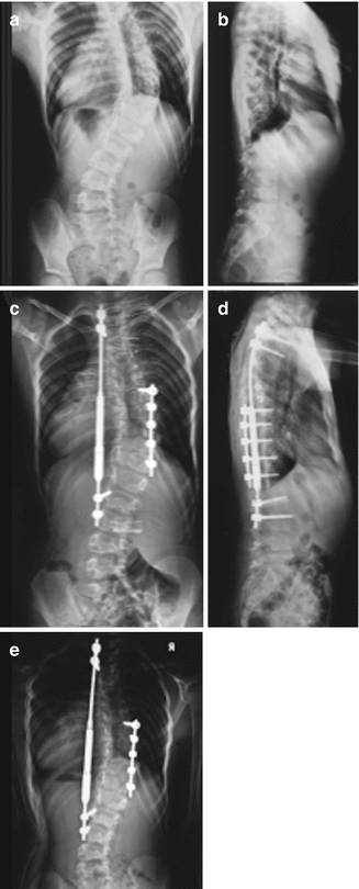
The techniques that employ convex hemiepiphysiodesis depend on the growth potential of the concave side of the anomalous segment for further correction and growth. However, this potential may be very small in congenitally abnormal vertebrae. In order to achieve a better trunk balance and progressive correction, we added a concave instrumented distraction construct to convex instrumented compression and fusion [1], similar to that suggested by Cheung et al. [6]. The initial results of this latest modification showed that the technique may provide immediate deformity correction, trunk balance, and continued growth of the spine (Fig. 40.3a–f). Currently, longer follow-up of the technique with a larger cohort of patients has become available [9]. The initial coronal deformity was 60.5° and corrected to 40.7° postoperatively. Distraction of the concave growing rod was performed every 6 months, and at a mean follow-up of 31 months, the final mean curve magnitude showed further correction at 35.5°. The sagittal plane was minimally affected from the procedure. Longitudinal growth of the T1–T12 segment has been noted in all patients in parallel with the improvement in deformity of mean 6.4 mm/year. Although our patients had multiple vertebral anomalies, the T1–T12 growth was only slightly less than the normative data published by Dimeglio for this age group [10]. This maybe due to the previously suggested growth stimulation by recurrent distractions of the growing construct [26].
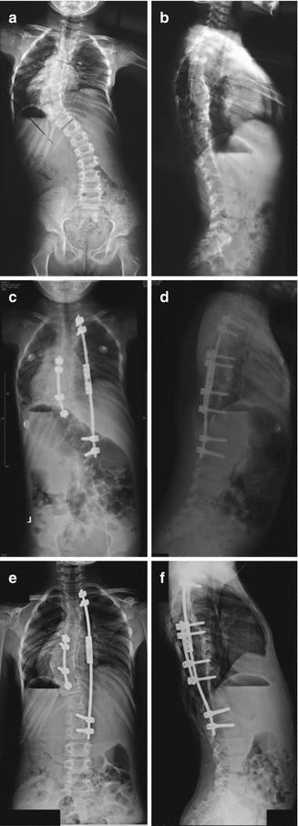
Fig. 40.3
A four-year-old female with (a) 60° thoracic scoliosis and coronal off balance and (b) hypokyphotic sagittal alignment underwent an instrumented convex growth arrest with concave distraction. Postoperative radiographs show that (c) coronal balance was achieved with substantial curve correction (40°), and (d) there appears to be a positive sagittal balance. At 2-year follow-up, the patient showed (e) 2 cm growth of the T1-S1 segment of the spine and further correction of the coronal deformity to 30°. The sagittal balance and alignment seems minimally changed from the preoperative radiograph (f)
The instrumented CGA with concave distraction appears to be more effective than the previously reported techniques of CGA, as there was no progression in the curve size but there was correction in all patients. The rationale for this technique is that the pedicle screws control the growth of the anomalous vertebral segments in the longitudinal [19] and transverse planes [7], and thereby obviating the need for an anterior fusion, while permitting spinal growth on the concave side of the curve through growth stimulation as a result of distraction [5]. Control of the whole curve by the concave growing rod also helped obtain immediate correction of coronal plane balance problems when compared with uninstrumented CGA. The potential advantages of this modification are that instrumentation on the convex side provides a complete hemiepiphysiodesis at the anterior and posterior convex sides, obviating the need for anterior surgery and enabling compression-rotation maneuvers for initial acute correction. Also, the instrumentation makes the procedure reliable by preventing potential inconsistencies that might arise from surgeon-dependent factors such as amount of end plate preparation.
One of the main disadvantages of the technique is that recurrent trips to the operating room are required for distraction (lengthening). This disadvantage may be circumvented by the utilization of the new magnetically controlled growing rod (MCGR). Our first trials with this technique show promise; however, the follow-up period is too short to make any solid conclusions (Fig. 40.4a–e). Another disadvantage arises from the use of instrumented hemiepiphysiodesis (hemifusion) of the apical vertebrae. Growing rod treatment is an alternative method for treatment of young children with a long curve and with a relatively flexible apical deformity including congenitally deformed vertebrae [12, 42]. However, growing rods do not control the apex of congenital curves with stiff anomalous segments involving more than four vertebrae, as were the typical case samples in the current study [42]. Another option for rigid, long sweeping congenital curves may be vertebral column resection [20] with limited posterior fusion. However, for such curves involving multiple anomalous segments, this technique causes shortening of the thoracic spine and necessitates the fusion of at least four to six additional thoracic levels for fixation after resection of the anomalous segments, thus interfering with thoracic growth. Moreover, this procedure is technically difficult and carries more neurologic risk compared with less complex procedures. Therefore, a less invasive method that preserves growth is warranted, whenever possible.
 Anesthesia and Postoperative Management of Spinal Deformity Surgery in Growing Children
Regulatory Policies Regarding Pediatric Spinal Devices
Imaging of the Growing Spine
Management of Spine Tumors in Young Children
Growth Modulation Techniques: Titanium Clip-Screw Implant System (HemiBridge)
Single and Dual Traditional Growing Rods
Anesthesia and Postoperative Management of Spinal Deformity Surgery in Growing Children
Regulatory Policies Regarding Pediatric Spinal Devices
Imaging of the Growing Spine
Management of Spine Tumors in Young Children
Growth Modulation Techniques: Titanium Clip-Screw Implant System (HemiBridge)
Single and Dual Traditional Growing Rods


Related posts:
Anesthesia and Postoperative Management of Spinal Deformity Surgery in Growing Children
Regulatory Policies Regarding Pediatric Spinal Devices
Imaging of the Growing Spine
Management of Spine Tumors in Young Children
Growth Modulation Techniques: Titanium Clip-Screw Implant System (HemiBridge)
Single and Dual Traditional Growing Rods
Stay updated, free articles. Join our Telegram channel
Full access? Get Clinical Tree
