Fig. 41.1
(a, b) The Shilla growing screw: a polyaxial pedicle screw with a cap that fixes to the top of the screw and snaps off for a low profile (b) and allows for the rod to be captured and able to slide in the screw
41.4.2 Surgical Technique
41.4.2.1 Preoperative Planning
Preoperative patient planning consists of a careful assessment of the upright coronal and sagittal films coupled with analysis of the flexibility of the curve via supine bend films, fulcrum bend films, or traction films. Determination of the location of the apical vertebral segments is of key importance. Those apical three or four vertebral segments that are least corrected through flexibility testing are the ones that comprise the apical levels for fusion and maximum correction. The goal is to render these segments neutral in all planes. If the surgeon can achieve this through posterior techniques alone (Ponte osteotomies, pedicle subtraction, or vertebral column resection), then no anterior release is necessary. For very stiff curves, anterior disk and end plate excision of the interval levels may be necessary before the planned correction. The posterior placed fixation at the apex uses bilateral fixed head pedicle screws. The Shilla growing screws are placed above (cephalad) and below (caudad) the apex to guide the growth of the spine at the ends of the curve and maintain coronal and sagittal correction. These screws are placed through the muscle layer without taking down soft tissues except to cut the fascial planes on each side of the midline. The use of a blueprint based on preoperative planning is helpful for the operating room team (Fig. 41.2).
Fig. 41.2
Example of a blueprint planned preoperatively and placed in the OR where the operating room team can refer to it during surgery
A single midline incision has been used to approach the three areas of instrumentation. Before incision of the fascia, radiographs are taken with small needle markers placed in the spinous processes to identify levels. Subperiosteal dissection is isolated to the apical levels only.
The fascia is incised one centimeter off the midline on both sides of the spinous processes from cephalad to caudad merging with the subperiosteal dissection at the apex. With direct visualization and either a freehanded technique or using C-arm fluoroscopy, bilateral pedicle screws are placed throughout the apical levels with fixed head screws. If an apical thoracoplasty is being used to enhance correction of the rib hump or for bone graft harvesting, it can be accomplished from the midline incision through the paraspinal muscle layers, removing the medial 2 cm from the apical deformed ribs.
Ponte osteotomies are performed between the apical segments and will enhance correction in all planes. Apical decortication will be necessary for fusion of these levels.
The Shilla growth guidance screws are placed through the muscular layer without visualization of the bone except radiographically (Fig. 41.3a–f). A cannulated polyaxial screw of sufficient diameter to fill the pedicle is used. The location of the Shilla screws is curve dependent but should extend into the lumbar spine sufficient to control the lordosis and the coronal curve. Avoid stopping the caudal instrumentation at the thoracolumbar junction. The Shilla screws can be placed at bilateral locations or staggered but should be separated apart a sufficient distance on the rod to allow for sliding of the rod easily. The Shilla screws at the top of the construct are subject to pull out forces from kyphosis and are best protected with a sublaminar wire placed one level above the upper screws. The wire can be placed with minimal dissection leaving the inner spinous ligament intact and removing the ligamentum flavum with a small Kerrison rongeur. The double wire can be split after sublaminar passage and passed through the soft tissues to each side without lifting the periosteum. Fiberwire (5 mm) is a good alternative to wire (Fig. 41.4).
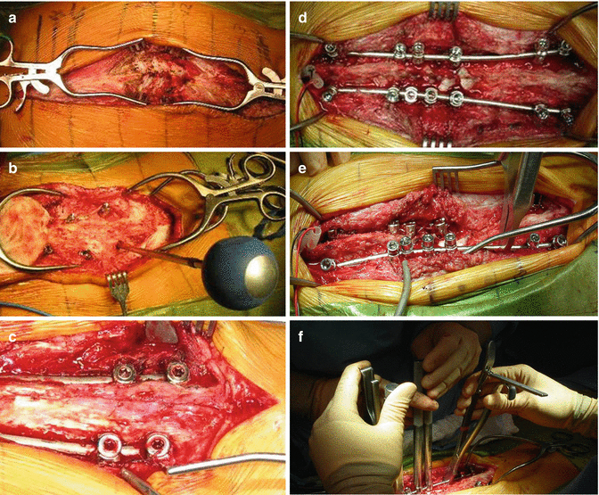
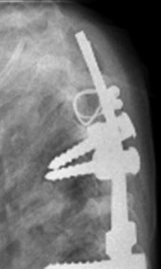
Fig. 41.3
(a–f) (a) A single incision is used, with levels identified using the C-arm, followed by subperiosteal dissection at the apex (cephalad on left, caudad on right). (b) Shilla screws are placed through the muscle. (c) And with C-arm guidance. (d) The rods are placed and rotated into the place with normal sagittal contours prebent into the rod. (e) Both rods in place. (f) Apical derotation with tube derotation
Fig. 41.4
Sublaminar wires can be placed cephalad above the Shilla screws to enhance fixation
The rod diameter is chosen appropriate for the size of the child. For smaller-sized children, the 4.5 mm rods are satisfactory; smaller diameter rods will break prematurely. Larger children (greater than 30 kg.) can tolerate the 5.5 mm rods which are more resistant to stress fractures in the metal. The 4.5 rods generally last 4–6 years before fracturing; the 5.5 rods last longer. The rod is contoured with normal sagittal curves, and the rod is left one level long at each end for growth (Fig. 41.5a–d).
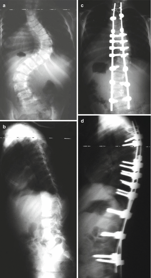
Fig. 41.5
(a–d) Preoperative (a, b) and postoperative (c, d) standing AP and lateral radiographs of a 4-year-old with Marfan’s and scoliosis treated with Shilla procedure
The apical levels are derotated with tube derotation devices or a vertebral column derotation device, while vice grips hold the rods in place to prevent rod rotation. The fixed head screws lock the rods at the apical screws via the locking set screws that press against the rods, while the Shilla caps capture the rods in the Shilla screws and press onto the polyaxial screw, not the rods. A cross-link is used to help counteract rod rotation. If the child is less than 5 years old, the cross-link should be avoided or a sliding type used to allow for growth in canal diameter. The torque/counter torque device snaps off the caps at a preset torque pressure. Bone graft is placed at the apex only. A small drain is often used.
Related posts:
 Anesthesia and Postoperative Management of Spinal Deformity Surgery in Growing Children
Regulatory Policies Regarding Pediatric Spinal Devices
Imaging of the Growing Spine
Orthotic Management for Early Onset Scoliosis
Growth Modulation Techniques: Titanium Clip-Screw Implant System (HemiBridge)
Single and Dual Traditional Growing Rods
Anesthesia and Postoperative Management of Spinal Deformity Surgery in Growing Children
Regulatory Policies Regarding Pediatric Spinal Devices
Imaging of the Growing Spine
Orthotic Management for Early Onset Scoliosis
Growth Modulation Techniques: Titanium Clip-Screw Implant System (HemiBridge)
Single and Dual Traditional Growing Rods
Stay updated, free articles. Join our Telegram channel

Full access? Get Clinical Tree


