Patterns of Unclear Significance


Patterns of Unclear Significance


Jonathan Charles Edwards

Ekrem Kutluay


The subject of patterns of unclear significance encompasses a selection of electroencephalographic findings that may look abnormal, but usually are not. These variants can be challenging, and may commonly lead to misinterpretation. These variants can be mainly grouped into two types: variant rhythms and variant transients. Some of these findings will also be discussed in the chapters on the normal EEG (Chapters 9 and 10).

The difficulties of interpretation posed by these variants arise for several reasons. These patterns possess features that are similar in some ways to abnormal patterns. Beginning with variant rhythms, as a general rule, a few common features help distinguish these rhythms from definitely abnormal findings: first, the variant rhythms are, for the most part, monomorphic. The term “monomorphic” simply means that each of the waves in the rhythm looks distinctly like the other waves in the rhythm. The waves have a characteristic shape, or “morphology,” that repeats. Second, most of the variants have an arch-shaped appearance, although some of the normal rhythms may have notches in some or all of the waves. Third, and most importantly, the variant rhythms for the most part do not evolve.

One important source of misinterpretation of these patterns may simply be that many of these patterns are uncommon. A significant number of clinicians who read EEGs in clinical practice may have only 2 to 3 months of closely supervised EEG training. The chances of seeing a rare pattern are quite low during this short training. Without extensive preparation in the fundamentals of electroencephalography and extensive supervised experience, misinterpretation of a rare pattern would not be surprising.

Finally, some confusion may arise from the fact that many of these patterns have changed names since first being described. In some cases, the nomenclature has changed numerous times. For some of patterns, numerous names are still concurrently in use today.

A particular challenge in assessing the true prevalence and clinical significance of these patterns is the tendency toward subject bias. Most patients who undergo EEG are doing so because of neurological symptoms, and many of these patients have a clinical history suggestive of epilepsy. While a few studies have looked at these findings in a general population, most have reviewed their presence in a clinical population. Since EEG is used to help diagnose epilepsy or other neurological conditions, the true predictive value of these findings may be biased (Tables 14-1 and 14-2).


FAST ALPHA VARIANT/SLOW ALPHA VARIANT

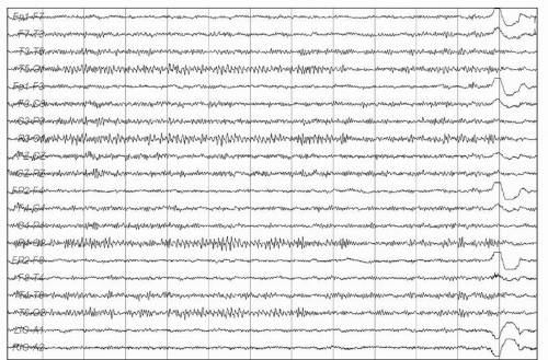
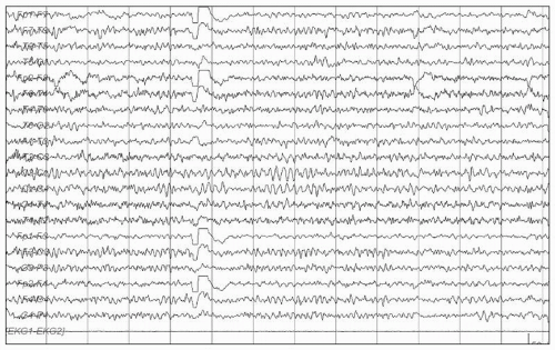
At times, harmonics or subharmonics of the background rhythm are seen. When this occurs, the resultant appearance is of a rhythm that is twice as fast or half as fast as the awake background. When the appearance is of a rhythm that is twice the frequency of the awake background, it is referred to as a “fast alpha variant” (1,2). A “slow alpha variant” has an apparent frequency that is about half that of the awake background (1, 2 and 3). These harmonics will often have a notched appearance, as if many of the waves are simply being cut in half, but only partly so. Slow and fast alpha variants are reactive to eye opening and closure (Fig. 14.1).

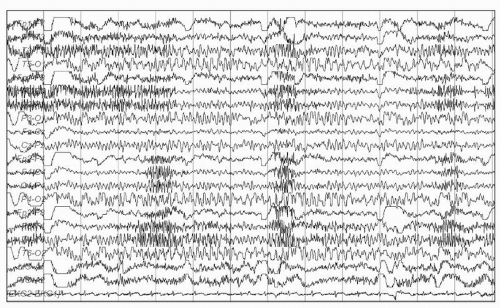
Fast alpha variant is easy to detect, since awake posterior background rhythms in the mid to upper beta range would be quite unusual. True beta rhythms are most often seen in the frontal, central, and parietal regions. When a posterior background rhythm of 16, 18, or 20 is seen, it is easy to think of fast alpha variant. However, slow alpha variant is more challenging, and may easily be misinterpreted as occipital slowing. This tendency to misread slow alpha variant as abnormal stems mostly from the fact that slowing is such an easily recognized abnormality, and all electroencephalographers look for signs of slowing. As a general helpful rule it is good to remember that any time one sees occipital slowing, one must think of slow alpha variant first, before calling the record abnormal. One should look to find a sample of normal awake background within the record. If the slowing is approximately half the frequency of the patient’s awake background, then the slowing is probably just a slow alpha variant. An additional clue can be small notches in some of the “slow waves,” bearing in mind that the notches can be quite subtle (Fig. 14.2).


ALPHA SQUEAK

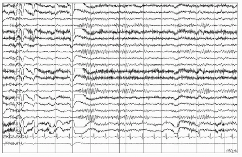
Immediately on eye closure, the awake background is sometimes initially faster, and of lower voltage. Over approximately 0.5 to 2 seconds, the background slows, and increases in amplitude to become the normal awake background. This initial “speeding up” of the background immediately on eye closure is referred to as an “alpha squeak,” and is a normal finding (Fig. 14.3) (2,4, 5 and 6).


Table 14.1 Summary of Characteristics for Patterns of Unclear Significance


Finding


Peak Age


State


Location


Frequency


Duration


Characteristics


Slow or fast alpha variant


Children and adults


Awake, eyes closed


Occipital


Half or double the posterior dominant rhythm


Brief or lasting many seconds


Sinusoidal, archiform, notched, flat-topped


Alpha squeak


Children and adults


Awake, immediately on eye closure


Occipital


Initially fast (beta) slowing rapidly to the posterior dominant rhythm


Typically 0.5-2 seconds


Sinusoidal, spindle-shaped


Rhythmic of midtemporal theta bursts of drowsiness (“RMTD”)


Younger to middle-aged adults


Drowsiness, light sleep


Midtemporal, at times with spread to parasagittal or occipital-temporal region
Bilateral or independent over the right or left side


5-7 Hz


Several seconds, or up to a minute or more


Arched, notched, flat-topped
Notche may give the waves a sharply contoured appearance


Midline theta rhythm (“Cigánek rhythm”)


Children and adults


Common in drowsiness and during mental activation. Less common in resting wakefulness


Midline (CZ, FZ), parasagittal


4-7 Hz


Brief or lasting many seconds


Arched, notched, flat-topped


Subclinical rhythmic electrographic discharge in adults (SREDA)


Adults, mostly age 40s-80s (mean 60s)


Drowsiness and resting wakefulness (rare cases reported in sleep)


Maximal in temporal and parietal regions
Usually bilateral, but may be asymmetric


May begin with delta frequency, but then increases to 4-7 Hz


Brief or lasting minutes


Sharply contoured, archiform, notched, flat-topped


14- and 6-Hz positive bursts


Childhood and adolescence


Drowsiness, light sleep


Posterior temporal


14 or 6 Hz


0.5-1 second


Archiform, comb-like, low amplitude


6-Hz spike and wave bursts


Children and adults


Drowsiness, light sleep


Highest amplitude over fronto-central regions


5-6 Hz


1-2 seconds


Usually low-amplitude (<25 µV) and short-duration (<30 msec) spike component


Benign sporadic sleep spikes


Mainly in adults. Also reported in children and adolescents


Drowsiness, light sleep


Anterior and midtemporal


Sporadic


Usually < 50 msec


Mono- or diphasic spike with steep descending arm


Wickets


Adults


Awake, sleep


Temporal regions


6-11 Hz if they occur in runs


Average of 2-4-second bursts in sleep


Archiform, monophasic


Frontal arousal rhythm


Children


Awakening, arousal from sleep


Frontal


6.5-8.5 Hz


Up to 13 seconds


Monomorphic


Table 14.2 Nomenclature Over Several Decades


EEG Pattern


Other Terminologies Used Previously


Rhythmic midtemporal theta bursts of drowsiness (RMTD)


Psychomotor variant (7)
Rhythmic midtemporal discharge (8)
Rhythmic theta bursts of drowsiness (1)
Rhythmic temporal theta bursts of drowsiness


Midline theta rhythm


Theta-discharges in the middle line (15)
Electrographic theta discharges in the midline (18)
Cigánek rhythm
Rhythmical midline theta (23)


Subclinical rhythmic EEG discharge of adults (SREDA)


Paroxysmal discharge of the temporo-parieto-occipital junction (24)


14- and 6-Hz positive bursts


14- and 6-Hz positive spikes (32), Ctenoids (33)


6-Hz spike and wave bursts


Spike and wave phantom (43)


Benign sporadic sleep spikes


Small sharp spikes (31), Benign epileptiform transients of sleep (50)


Wickets


Wicket spikes (58)


Frontal arousal rhythm


Frontal arousal rhythm (63)


RHYTHMIC MIDTEMPORAL THETA BURSTS OF DROWSINESS

First described by Gibbs and Gibbs, this pattern has been given several names over nearly six decades (7). This pattern’s earlier name “psychomotor variant” is still commonly used. “Psychomotor” is an older term for a complex partial seizure, typically of temporal lobe origin. Rhythmic midtemporal theta bursts of drowsiness (RMTD) has commonly been referred to as “psychomotor variant,” because it was felt to have a strong resemblance to a temporal lobe seizure. Subsequent names have included: “rhythmic midtemporal discharge” (8), “rhythmic theta bursts of drowsiness,” and “rhythmic temporal theta bursts of drowsiness” (1).

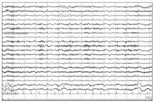
The name RMTD is widely accepted, and quite clear in listing the most distinctive characteristics. RMTD is seen mostly during drowsiness and light sleep, although it may rarely be seen during wakefulness. It occurs in bursts or trains of rhythmic 5- to 7-Hz activity. RMTD is maximal in the midtemporal leads, and may at times spread to the parasagittal regions, or more posteriorly to the occipital-temporal region. Like mu rhythm, RMTD may be seen bilaterally or may be independent over the right or left side. Like most rhythmic variants, it is typically arch-shaped, and is often notched. These notches may give the waves a somewhat sharply contoured appearance, which may be mistaken for a sharp wave. At times, the notched arches may have a flat-topped appearance (1,7,8). RMTD may occur in runs that last several seconds, or even up to a minute or more. However, like other normal rhythms, RMTD can be clearly distinguished from seizure activity, because it is monomorphic, monorhythmic, and does not evolve.

The reported overall prevalence of RMTD is quite low, ranging from 0.1% to 2% of records (9, 10, 11, 12 and 13). The prevalence appears to be lower in age-matched normal controls than it is in EEGs performed in neurological patients (9). While some studies (8) have reported that a significant number of patients who are found to have RMTD may have epilepsy, the majority of patients with this finding will not. RMTD has been reported in the presence of a structural abnormality (14). The overall clinical significance of this finding is unclear. It is most commonly considered a normal variant (Fig. 14.4).


MIDLINE THETA RHYTHM (CIGÁNEK RHYTHM)

Midline theta rhythm was first described by Cigánek, and is still commonly referred to as “Cigánek rhythm” (15). As is the case with many variant rhythms, the midline theta rhythm was originally thought to be indicative of epilepsy, but was later found to be present in normal subjects also. Cigánek’s 1961 report is actually titled “Theta-discharges in the middle line—EEG symptom of temporal lobe epilepsy.” Midline theta is seen during wakefulness and drowsiness. Similar to RMTD, the midline theta rhythm is typically a 4- to 7-Hz arch-shaped or sinusoidal rhythm that may at times be notched or flat-topped (16). The main difference in appearance from RMTD is the location (typically maximum over CZ), and presence in wakefulness.

The reported prevalence of midline theta rhythm has been highly variable, ranging from 2% to 35% of subjects (17,18). This wide disparity is in part accounted for by subject selection. Maulsby included the presence of midline theta during drowsiness and determined that it was present in 35% of normal adults. Many others have reported midline theta during mental tasks in a high number of patients (19, 20, 21 and 22). With the exclusion of midline theta rhythm that is seen only during drowsiness of mental activation, the prevalence has been reported to be low in normal controls, and higher in patients with epilepsy. A more recent study (23) suggested that when drowsiness and mental activation are excluded, midline theta rhythm was common (26% overall) in their patients with epilepsy, and uncommon (0/54) in controls. Interestingly, in the same series, and using the same exclusion criteria, midline theta rhythm was seen much more frequently in frontal lobe epilepsy (48.1%) than in temporal lobe epilepsy (3.7%).

The clinical significance of midline theta rhythm is not universally agreed upon (16,23). It is typically regarded as a non specific finding, and is generally read as normal. Midline theta rhythm during the awake recording and without mental tasks may
warrant further study. A potential practical challenge and confounding variable would be in actually knowing what a subject is doing mentally in an awake, at rest recording, and ruling out that particular mental tasks are taking place. Most evidence suggests that the presence of midline theta rhythm during drowsiness or during mental task is not an abnormality (Fig. 14.5).


Figure 14.1 Fast alpha variant.


Figure 14.2 Slow alpha variant.


Figure 14.3 Alpha squeak.


Figure 14.4 Rhythmic midtemporal theta bursts of drowsiness.


Figure 14.5 Midline theta rhythm.


SREDA (SUBCLINICAL RHYTHMIC ELECTROGRAPHIC DISCHARGE IN ADULTS)

SREDA is a fairly rare pattern that can be easily misinterpreted. It is perhaps the most challenging pattern among those that are considered normal variants. First described in 1961 by Naquet et al., this pattern was originally thought to be associated with hypoxia (24). Westmoreland and Klass described 65 patients who had 142 EEG recordings, and provided the term “Subclinical Rhythmic EEG Discharge in Adults.” In this series, SREDA was seen in patients aged 42 to 80, with an average age of 61 (25).

Only gold members can continue reading. Log In or Register to continue

Stay updated, free articles. Join our Telegram channel

Sep 9, 2016 | Posted by in NEUROSURGERY | Comments Off on Patterns of Unclear Significance

Full access? Get Clinical Tree

Get Clinical Tree app for offline access