Pediatric Neuroimmunology


Pediatric Neuroimmunology


Leslie A. Benson

Riley Bove

Mark Gorman


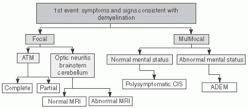
DEMYELINATING DISEASES (see Fig. 17.1 and Table 17.1)


FIGURE 17.1 Flow Chart Demonstrates Approach to a First Demyelinating Syndrome Based on Focality of the Signs and Symptoms. CIS, clinically isolated syndrome. MRI is generally the first diagnostic testing choice to help categorize inflammatory etiologies. ATM, acute transverse myelitis; ADEM, acute disseminated encephalomyelitis.


TABLE 17.1 Major Causes of White Matter Changes and CNS Inflammation


Autoimmune


Primary demyelinating


ON, ADEM, TM, CIS, MS (RRMS, SPMS, PPMS), NMO


Paraneoplastic


Paraneoplastic encephalomyelitis, OMS


Connective tissue dz


SLE, Behçet disease, RA, Sjögren syndrome, APLAS, HLH


Granulomatous


Wegener granulomatosis, sarcoidosis, lymphomatoid granulomatosis


Vasculitis


CNS and systemic vasculitides


Other Demyelinating


Infectious


CMV, HIV → PML (JCV, usually in HIV), Lyme, neurosyphilis, HTLV1, SSPE, tropical spastic paraparesis/HTLV1-assoc. myelopathy, congenital toxoplasmosis


Metabolic


CPM, subacute combined degeneration (B12 & intrinsic factor deficiency)


Toxic


NO poisoning; drugs of abuse (toluene, ethanol, cocaine, MDMA/ecstasy, IV/inhaled heroin, psilocybin); radiation-induced necrosis; anti-neoplastic agents (methotrexate, carmustine, cisplatin, cytarabine, fluorouracil, levamisole, fludarabine, thiotepa, interleukin-2, interferon-α); immunosuppressants (Cyclosporine [posterior lobes], tacrolimus [parieto-occipital]); antimicrobials (amphotericin B, hexachlorophene, metronidazole); environmental exposure (carbon monoxide, arsenic, carbon tetrachloride).


Vascular & hypoxic-ischemic


Migraine, PRES, HIE, sickle cell disease, delayed hypoxic cerebral demyelination, CADASIL


Secondary demyelinating


X-linked adrenoleukodystrophy, metachromatic leukodystrophy, adrenomyeloneuropathy, Alexander, Canavan, Krabbe, Aicardi-Goutières syndrome


Hypomyelinating


Pelizaeus-Merzbacher, Cockayne syndrome


Mitochondrial disease


NARP, POLG1/Alpers, MELAS, Leigh syndrome


Neoplasm


Glioma, lymphoma, LCH


Note: Most common in bold.


ADEM, acute disseminated encephalomyelopathy; APLAS, antiphospholipid antibody syndrome; CADASIL, cerebral autosomal dominant arteriopathy with subcortical infarcts and leukoencephalopathy; CIS, clinical isolated syndrome; CPM, central pontine myelinolysis; HIE, hypoxic ischemic encephalopathy; HLH, hemophagocytic lymphohistiocytosis/macrophage activation syndrome; JCV, JC virus; LCH, Langerhans cell histiocytosis; MELAS, multifocal encephalopathy lactic acidosis and strokes; MS, multiple sclerosis; NARP, neuropathy ataxia and retinitis pigmentosa; NMO, neuromyelitis optica; NO, nitric oxide; OMS, opsoclonus-myoclonus syndrome; ON, optic neuritis; PML, progressive multifocal leukoencephalopathy; POLG1, polymerase gamma 1; PPMS, persistently progressive MS; PRES, posterior reversible encephalopathy syndrome; RA, rheumatoid arthritis; RRMS, rapidly progressive MS; SLE, systemic lupus erythematosus; SPMS, secondary progressive MS; SSPE, subacute sclerosing panencephalitis; TM, transverse myelitis.


VACCINE CONSIDERATIONS: Empiric recommendation for no vaccines for 1 mo after Clinically isolated syndrome (CIS)/MS, 3 mo for acute disseminated encephalomyelitis (ADEM). Consult Red Book for patients on steroids, IVIG, and timmunosuppressants.


Acute Disseminated Encephalomyelitis (ADEM)

DEFINITION: A first clinical event with a polysymptomatic presentation including encephalopathy, with acute or subacute onset.

EPIDEMIOLOGY: 0.4 to 0.8 per 100,000 incidence in children & adolescents (average age 5-8 y); rare in adults.1,2

ETIOLOGY: Postinfectious: ˜75%; Postvaccination: ˜5%; Usually after MMR, but risk is greater and disease more severe w/ measles (1/1,000) and rubella (1/5,000) than MMR vaccine (1-2/million). Others: Rabies, HBV, influenza, DPT, Japanese B encephalitis, pneumococcal, polio, and rare cases in organ transplant patients. Typically within 4 to 14 d of infection or vaccination; must be <3 mo to be associated.

PATHOLOGY: Perivenular T-cell and macrophage infiltrates associated with demyelination.


PATHOPHYSIOLOGY: Proposed immune dysregulation in genetically susceptible individuals triggered by environmental stimulus. Anti-myelin oligodendrocytes (MOG) antibodies detected in some patients are of uncertain significance.


Diagnostic criteria3


  • Clinical Features: (1) First clinical attack of inflammatory or demyelinating dz in CNS. (2) Acute or subacute onset ( febrile illness <4 wk prior in 50%-75% children; max severity over 4-7 d). (3) Polyfocal: affects multiple areas of CNS. (4) Polysymptomatic (e.g., n/v, meningismus, cranial neuropathies including optic neuritis (ON); pyramidal signs, hemiparesis, ataxia, transverse myelitis (TM), seizure (in ˜1/3) (5) Must include encephalopathy: Acute behavioral Δ (e.g., confusion or irritability) and/or alteration in consciousness (range: somnolence-coma). (6) Subsequent improvement by clinical/imaging measures (but may have residual deficits). (7) No better explanation (other etiologies ruled out). (8) Relapses: new or fluctuating signs or MRI findings within 3 mo of inciting ADEM episode are considered part of the initial event.


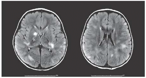
  • Lesion Characteristics on MRI FLAIR and T2: (1) Large (>1-2 cm) multifocal, hyperintense, bilateral, asymmetric lesions in supratentorial or infratentorial WM, <50% of patients have contrast enhancement (Fig. 17.2). Rarely: single large lesion. (2) May involve basal ganglia and thalamus. (3) Spinal cord may show confluent intramedullary lesions. (4) No radiologic evidence of prior destructive WM changes, e.g., no black holes (most ADEM lesions resolve at least partially vs. MS lesions).


Diagnostic testing


  • MRI w/ T2, FLAIR, and gadolinium. Sequential imaging to evaluate. If new symptoms, consider multiphasic ADEM vs. MS.


  • LP: r/o viral and bacterial meningitis/encephalitis. Send CSF for: protein, glucose, cell count, Gram stain, bacterial culture, and oligoclonal bands (OCB). Consider further infectious testing based on immune status, clinical scenario, CSF WBC count. CSF can be normal. May see pleocytosis and/or increased protein, OCB in <10% (nonspecific—also seen in MS, CNS infections).


  • Serum: CBC; may have increased ESR and CRP.


  • Micro: Studies negative.


  • EEG: Background slow wave activity c/w encephalopathy.


FIGURE 17.2 Typical MRI Features in ADEM. FLAIR sequences demonstrating multifocal fluffy, bilateral, asymmetric hyperintensities in the bilateral white matter and deep gray nuclei.


DIFFERENTIAL DIAGNOSIS: (1) Infectious: Viral/bacterial meningitis or encephalitis. (2) Inflammatory demyelinating d/os: CIS (encephalopathy required for ADEM dx, not typical for CIS/MS. CSF >50 WBC/mm, sometimes seen in ADEM, atypical for MS. MRI shows old lesions for MS), ON, TM, neuromyelitis optica (NMO), and others. (3) CNS vasculitis (PACNS; see below) (4) MalignancyCNS lymphoma or glioma; workup with CSF cytology but dx requires brain biopsy. (5) Mitochondrial disease—w/ intercurrent infection. MRI w/ typical symmetric T2 signal in basal ganglia or parieto-occipital regions and lactate peak on MR SPECT; elevated serum and CSF lactate.

TREATMENT: No controlled trials to date. (1) Antibiotics: Consider empiric acyclovir & antibiotics until infectious etiology excluded. (2) Steroids: IV methylprednisolone 30 mg/kg/d (max 1 g/d) then 3- to 6-wk oral prednisone taper (+ PPI, Ca/vitamin D). Two studies suggest less early recurrences with taper >3 wk. (3) Steroid alternatives: If steroids fail or contraindicated, can use plasmapheresis (PLEX), IVIG.3a,3b

PROGNOSIS: Overall good: ˜75% recover completely, <5% mortality. MRI can show complete recovery. Average recovery time of 1 to 6 mo. 3.3% with ADEM younger than 11.85 y develop MS.4 Motor deficits: 8% to 30% residual; range from clumsiness to ataxia and hemiparesis. Neurocognitive deficits: attention, short-term memory, affect, behavior, verbal processing speed, but less severe than in MS, usually within 1 standard deviation from norm. Poor prognostic factors: adults, unresponsive to steroids.

ADEM VARIANTS: Multiphasic ADEM: New event meeting the criteria for ADEM 3 or more months after initial ADEM event. Acute hemorrhagic leukoencephalitis: Includes AHL—acute hemorrhagic leukoencephalitis, AHEM—acute hemorrhagic encephalomyelitis, and ANHLE—acute necrotizing hemorrhagic leukoencephalitis (Weston Hurst syndrome). A hyperacute variant with necrotizing vasculitis (venules) w/ hemorrhage and edema, very rare (2% of ADEM), poorer prognosis (70% mortality usually within 1st wk; 70% survivors w/ residual deficits). Prognosis may be improved w/ aggressive treatment (corticosteroids, IVIG, cyclophosphamide, plasmapheresis). Most cases occur after URI. MRI: diffuse WM lesions within 72 h of onset, cerebral edema, ICH. CSF: increased WBC, RBCS, and protein.


Clinically Isolated Syndrome/Acute Demyelinating Syndrome (ADS)3: Isolated event of CNS demyelination

DEFINITIONS: Polyfocal—clinical features attributed to more than one CNS site without encephalopathy. Monofocal—clinical features attributed to only one CNS site (i.e., optic nerve, spinal cord, cerebrum, brainstem or cerebellum).

EPIDEMIOLOGY: ADS incidence 0.9/100,000 children in Canada (including ADEM).5

DIAGNOSTIC TESTING: Aimed at ruling out alternative diagnoses & evaluating risk of MS.


  • MRI brain w/ and without gad: Include orbits if suspect ON. Consider spine in all cases.


  • LP: Routine (protein, glucose, cell count/diff, Gram stain, and culture); IgG index, OCB (if present, increase risk of subsequent MS). Usual findings: mild pleocytosis, elevated protein, or OCBs.


  • Labs: CBC (mild elevation in WBC in 50%), ESR, CRP, ANA, Lyme, EBV titers.


  • MANAGEMENT: Follow-up imaging brain/spine MRI at 1 to 3 and 6 mo, then 6 to 12 mo depending on suspicion for dissemination, looking for new lesions and fulfillment of McDonald criteria (see MS diagnosis below).


PROGNOSIS: Risk of MS lower in children than adults. After monofocal CIS, 38% to 62% vs. 0% to 17% with ADEM developed MS.6 Poor prognostic indicators include age >10 to 12 y, asymptomatic MRI brain lesions (strongest predictor), CSF OCB, HLA-DRB1*15, prior EBV infection, low 25-hydroxy vitamin D. MS risk after ADS is 1.9% w/ normal brain MRI, 28% with abnormal MRI in patients ≤11.85 y, and 60.6% with abnormal MRI in patients ≥11.85 y.4


Optic Neuritis (ON)

DEFINITION: Inflammatory demyelination of the optic nerve, causing acute visual loss. Can occur along any segment; termed retrobulbar if posterior to the optic nerve head. Common manifestation of MS.

EPIDEMIOLOGY: 1:1.4 female:male.5 May be para-infectious.

CLINICAL FEATURES: Acute: (1) Binocular (more common in children than adults) or monocular visual loss over hours to days, peaking in 1 to 2 wk. (2) Loss of color vision—test for red desaturation. (3) Visual field defect—usually central scotoma. (4) Eye pain (56%)7 in the orbital or retroorbital region usually exacerbated w/ eye movement, which often precedes visual loss. Chronic: Uhthoff phenomenon: heat (showers, exercise) increases visual problems, which resolve when cooled off. Associations: MS, NMO, ADEM.

DIAGNOSTIC TESTING: A clinical diagnosis, but confirmatory testing is often necessary in children. In addition to CIS workup, consult ophthalmology, preferably neuro-ophthalmology.


  • Visual acuity (VA) testing: Median VA <20/200 at peak in 70% of children.7,8,9 Persistent visual loss (color vision, contrast sensitivity, light brightness, stereo acuity, motion detection) detectable in most at 2 y.


  • Color testing: Ishihara plates or Farnsworth-Munsell 100 hue test can be used. Color vision loss in about 90% patients, out of proportion to decrease in VA. “Red” desaturation: appears “washed out” or darker in affected eye.


  • Flashlight test: Relative afferent pupillary defect in unilateral or asymmetric ON. It can persist.


  • Visual fields: Usually central scotoma. If extends to the periphery, consider compressive lesion.


  • Fundoscopic examination: Papillitis w/ hyperemia and disk swelling, blurred disk margins, distended veins (60%-72% in pediatrics).9,10 Retrobulbar neuritis w/ normal fundoscopic exam.


  • Over time optic atrophy occurs, even if VA is normal, particularly temporal, extending beyond its margin into peripapillary.


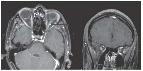
  • MRI brain and orbits w/ and w/o gad: Can have optic nerve inflammation with T2 bright signal w/ or w/o enhancement (T2 signal change best seen on coronal STIR) but normal does not r/o dx (Fig. 17.3). Brain MRI for prognosis (see below).


  • LP: Role in isolated ON controversial. If brain parenchymal lesions, LP can help stratify MS risk.


  • Labs: See CIS; consider NMO IgG in severe and/or bilateral cases.11


  • Visual evoked potential (VEP): Consider if diagnosis not clear; will show decreased amplitude of N95 & delay in the P100.


  • Optical coherence tomography (OCT): Detects thinning of retinal nerve fiber layer once early swelling is gone.

DIFFERENTIAL DIAGNOSIS: (1) Infectious neuroretinitis (viruses, toxoplasmosis, Bartonella) often bilateral, ± scotoma, ± pain. Exam: papillitis, macular edema, exudates. ± systemic signs/symptoms. (2) NMO.


FIGURE 17.3 Typical Optic Neuritis. MRI orbits, T1—post-gad axial and coronal images demonstrate enhancement within the optic nerve (arrows) on the left (compared to the normal right nerve).

(3) Genetic: LHON: in boys with onset over weeks to months, can be unilateral or bilateral with central or cecocentral scotoma. Fundus with disc hyperemia; peripapillary telangiectasia. Not painful. MRI normal. Smoking assoc. w/ attack. (4) Inflammatory: Autoimmune (SLE, Sjögren), sarcoidosis. (5) Neoplasms: Optic nerve glioma—initial symptoms often identical. MRI with expansile enhancing mass of optic nerve. Can have proptosis and other orbital signs. Frequent in NF1. (6) Compressive lesion. (7) Pseudotumor cerebri—vision loss usually late and HA frequently present. (8) Chronic relapsing idiopathic optic neuropathy (CRION). (9) White dot syndrome. (10) Toxic-metabolic: Drugs, toxins, nutritional deficiency (vitamins B1, B12, folate), radiation. (11) Traumatic optic neuropathy. (12) Conversion disorder.

TREATMENT: Acute: No randomized clinical trials (RCTs). The ON treatment trial found more rapid improvement with IV steroids if VA <20/40 but no difference in long-term outcome. Most tend to treat. IV methylprednisolone (30 mg/kg up to 1 g IV daily ×3 to 5 d + PPI, Ca/vitamin D). PO prednisone alone is NOT recommended.12,13 If poor recovery, consider 2nd-line agent such as IVIG or PLEX. If refractory, consider NMO.

Only gold members can continue reading. Log In or Register to continue

Stay updated, free articles. Join our Telegram channel

Jun 20, 2016 | Posted by in NEUROLOGY | Comments Off on Pediatric Neuroimmunology

Full access? Get Clinical Tree

Get Clinical Tree app for offline access