42 Schwannomas are slowly growing neoplasms composed of Schwann cells. Solitary schwannomas with effects on the CNS occur on cranial (Table 42.1, Fig. 42.1) and spinal nerve roots; rarely they are found in the substance of the brain or spinal cord. Melanotic schwannomas have a predilection for spinal nerve roots. The plexiform schwannoma is rare and occurs in the skin. Unlike the plexiform neurofibroma, it is not associated with NF. Table 42.1 The commonest neoplasm of the cerebellopontine angle is a schwannoma of the vestibulocochlear nerve 42.1 Vestibulocochlear nerve schwannomas. Clinical criteria for diagnosing NF1 Two or more of the following clinical features must be present to fulfil current diagnostic criteria for NF1: Schwannomas are composed of nodular rubbery tissue, which has a variegated cut surface. Yellow and gray areas may be interspersed with hemorrhagic foci or cysts. The neoplasm has a capsule and the nerve from which the schwannoma arises may be splayed over the surface of the neoplasm (Figs 42.2, 42.3). 42.2 The relationship of neurofibroma and schwannoma to associated nerve. 42.3 The relationship between schwannoma and peripheral nerve. Two histologic patterns predominate (Figs 42.4–42.6): 42.5 Schwannoma. 42.6 Schwannoma.
Peripheral nerve sheath neoplasms
 GENETICS OF NEUROFIBROMATOSIS TYPE 1 (NF1)
SCHWANNOMAS
Cerebellopontine angle neoplasm
%
Schwannoma
85
Meningioma
10
Others (cholesteatoma, glioma, medulloblastoma, metastatic carcinoma, paraganglioma, hemangioma, AT/RT)
5
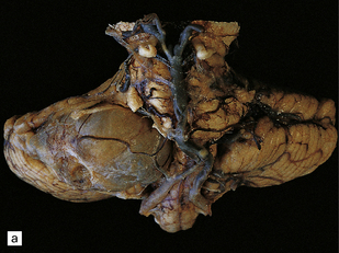
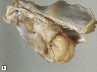
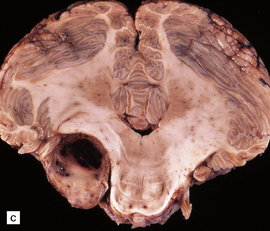
(a) A partly cystic schwannoma in the cerebellopontine angle indents the brain stem. Flattened fascicles of vestibulocochlear nerve run over the surface of the neoplasm. (b) A schwannoma sits at the mouth of the internal auditory meatus. (c) A hemorrhage occupies part of this schwannoma, which has caused compression and distortion of the brain stem.
 At least two solitary neurofibromas or one plexiform neurofibroma.
 At least two Lisch nodules (iris hamartomas).
 Optic glioma (pilocytic astrocytoma).
 Osseous lesions (sphenoid dysplasia, pseudoarthrosis, or spinal deformity).
 A family history of a first-degree relative with NF1 fulfilling the above criteria.
MACROSCOPIC APPEARANCES
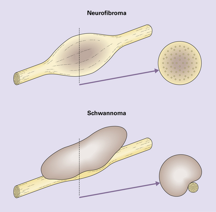
The neurofibroma incorporates axons, but the schwannoma displaces normal elements of the nerve to one side (see Fig. 42.3).
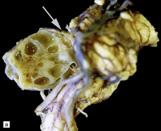
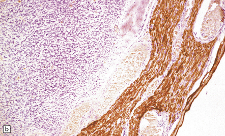
(a) A cystic schwannoma arising from the eighth nerve (arrow) distorts the brain stem. The tumor has been hemisected to reveal its multicystic architecture. (b) An antibody to neurofilament protein labels axons in a nerve alongside a schwannoma.
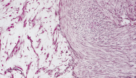
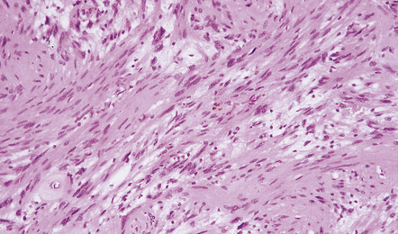
MICROSCOPIC APPEARANCES
Abundant pericellular reticulin in an Antoni A area. Section adjacent to that in Figure 42.4.
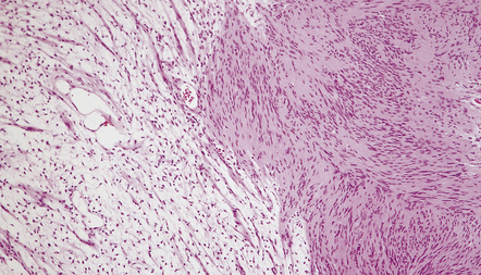
Fascicles of spindle-shaped cells are interrupted by small myxoid areas. Note the small blood vessel with a markedly thickened wall.
 Antoni A areas, in which spindle-shaped cells with rod-shaped nuclei and dense pericellular reticulin are arranged in compact intertwining fascicles.
 GENETIC ASPECTS OF NERVE SHEATH NEOPLASMS
 GENETICS OF NEUROFIBROMATOSIS TYPE 2 (NF2)


 NF1
NF1


 NF2
NF2


