Piriformis Syndrome, Obturator Internus Syndrome, Pudendal Nerve Entrapment, and Other Pelvic Entrapments

CHAPTER 238 Piriformis Syndrome, Obturator Internus Syndrome, Pudendal Nerve Entrapment, and Other Pelvic Entrapments


Nerve entrapments of the posterior pelvis (sciatic nerve, posterior femoral cutaneous nerve, superior gluteal nerve), inferior pelvis (pudendal nerve, nerve to the obturator internus, obturator nerve), and anterior pelvis (ilioinguinal nerve, femoral nerve, lateral femoral cutaneous nerves) and of other nerves of the pelvis are now increasingly well understood. Radiating pain in the low back, buttock, and leg is among the most common symptoms of patients seen by neurosurgeons. In addition to herniated lumbar disks, these symptoms may be caused by entrapment or irritation of several different peripheral nerves.


Superior gluteal nerve syndromes are relatively rare but should be considered in patients with unilateral pain involving the low back below the level of iliac crests in whom no clear relevant root impingement or ipsilateral facet abnormality can be located in the spine. In addition to the obvious upper buttock distribution of symptoms from pain and spasm in the gluteus maximus and gluteus medius, there may also be pain in the lateral thigh between the greater trochanter and the knee. This is because the tensor fascia lata muscle is also innervated by the superior gluteal nerve.


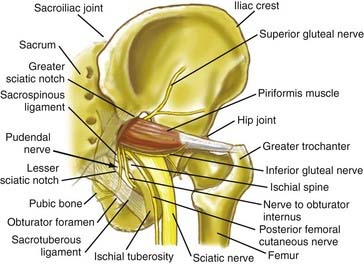
Pain in the upper buttock over the posterior superior iliac spine may also be due to entrapment of the superior cluneal nerves. Nonradiating pain in the mid buttock may be direct pain from the piriformis muscle or may be related to a trochanteric bursitis. Pain involving the piriformis muscle often includes associated radiating nerve pain such as sciatica because the sciatic nerve passes through the sciatic notch along with the piriformis muscle (Fig. 238-1).


The posterior femoral cutaneous nerve runs with the sciatic nerve from its exit through the sciatic notch and along their course together into the upper thigh. It has inferior cluneal branches that can be associated with low buttock pain, but its main distribution is along the posterior thigh. By contrast, the sciatic nerve can be responsible for deep pain in the hamstrings but is more commonly represented by tibial nerve pain in the posterior calf and sole of the foot and by peroneal nerve pain in the anterior portion of the lower leg and the top of the foot.


Pain in the medial thigh may be associated with entrapments of the obturator nerve. Pain in the anterolateral thigh may be from lateral femoral cutaneous nerve entrapment, meralgia paresthetica, typically reflecting entrapments near the anterior superior iliac spine. Only truly anterior thigh pain should be considered as femoral nerve distribution. Pain due to femoral nerve entrapment will typically extend to the medial ankle by consequence of distal involvement of the saphenous nerve—a descendant component of the femoral nerve.


Physical examination may reveal pain on the lateral aspect of the ischial tuberosity in patients who have sciatic nerve entrapments in the ischial tunnel. In the upper ischial tunnel, the sciatic nerve may be affected by the distal tendon of the obturator internus muscle. In the lower ischial tunnel, pathology affecting the sciatic nerve and posterior femoral cutaneous nerve is often associated with tears or irritation affecting the hamstring attachment on the ischial tuberosity. Just at and below the gluteal crease, there may be sciatic entrapment associated with the quadratus femoris muscle.


On the medial aspect of the ischial tuberosity, palpation can detect spasm in the obturator internus muscle. This is often experienced by the patient as sitting pain. It can also present as groin pain or hip pain. Hip pathology can be ruled out by tests of passive hip rotation and appropriate imaging studies such as hip radiographs and pelvis magnetic resonance imaging (MRI).


The neurosurgeon can palpate along the inguinal ligament to check for radiating pain from ilioinguinal and genitofemoral nerve entrapment. Obturator internus muscle pain is often detectable as sensitivity to palpation at the obturator foramen in patients without other inguinal sensitivity or inguinal hernia.


Spasm in the obturator internus muscle is most often caused by irritation or entrapment of the nerve to the obturator internus. This nerve exits the greater sciatic notch between the sciatic nerve and the pudendal nerve and then branches in the retrosciatic space, sending most of its descendant elements through the lesser sciatic notch to innervate the muscle.


Three types of radiating nerve symptoms can result from spasm of the obturator internus muscle. The spasm may impinge on the transiting obturator nerve, causing medial thigh and adductor symptoms. It may also impinge on the sciatic nerve where that nerve crosses the obturator internus tendon in the upper portion of the ischial tunnel. Most important, by flattening the entrance to Alcock’s canal, it can cause impingement of the pudendal nerve. Pudendal nerve entrapment syndromes present with pain and numbness in the genitalia and rectum and other saddle area distributions. They can also be associated with bladder dysfunction, pelvic floor pain, and sexual dysfunction. Unlike other causes of urogenital pain and dysfunction, these symptoms resolve, at least transiently, when the obturator internus muscle is relaxed by a bupivacaine (Marcaine) injection and resolve permanently with neuroplasty release of the nerve to the obturator internus and the pudendal nerve. Pudendal nerve entrapments can happen at the level of the greater sciatic notch in association with a piriformis muscle syndrome, at the level of ischial spine in association with the sacrotuberous and sacrospinous ligaments, and at the entrance or exit of Alcock’s canal.


Diagnosis and Management of Pelvic Sciatic Syndromes


Physical Examination Findings in Pelvic Sciatic Entrapment Syndromes


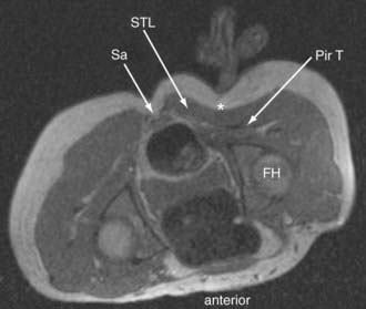
Sciatic entrapments at the sciatic notch often affect all five toes (multiple dermatomes) rather than just the lateral toes (S1 radiculopathy) or medial toes (L5 radiculopathy), which is most commonly seen in those with herniated lumbar disks. The pain of sciatic entrapment more commonly extends primarily only as far as the knee, ankle, or heel—not reaching the toes at all. Straight leg raising is generally negative, but resisted abduction or adduction of the flexed internally rotated thigh usually reproduces the symptoms. Sciatic notch tenderness is present in all patients with piriformis syndromes (Fig. 238-2), so other variant types of pelvic sciatic entrapment should be considered when this examination feature is not present. Trochanteric bursitis responsive to injection of the bursa occurred in 7% of patients diagnosed with muscle-based piriformis syndrome.


Patients with entrapments at the level of the ischial tuberosity (ischial tunnel syndrome) have tenderness to palpation at the lateral surface of the ischial tuberosity, which is about 4 inches below the level of the sciatic notch.


Neurography Results for Sciatica of Nondisk Origin


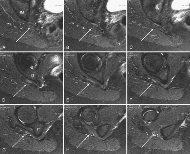
Until recently, objective tests for the existence of piriformis syndrome were very limited, there was no reliable effective treatment, and the pathophysiology was not well understood.1 Piriformis syndrome remains a subject of significant debate within the neurosurgical community.2,3 However, magnetic resonance neurography has proved helpful in providing objective diagnostic criteria.4 Figure 238-3 demonstrates the appearance of the sciatic nerve in a magnetic resonance neurography image in a typical pelvic sciatic entrapment case. Ipsilateral piriformis muscle hypertrophy is a common image finding in piriformis syndrome.5,6 Ipsilateral muscle atrophy occurs in some patients as well. Edema or hyperintensity in the ipsilateral sciatic nerve relative to the contralateral nerve occurs in 88%.4


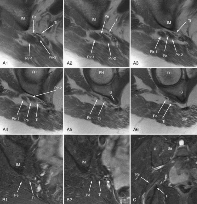
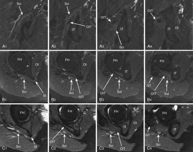
In addition to entrapment at the sciatic notch, the sciatic nerve may be compressed by fibrous bands and by dilated venous varices when the dilated vein arises inside the perineurial sheath. It may suffer entrapment owing to fixation by an artery passing through the nerve and by tendon or muscle passing through the nerve (Fig. 238-4). In some individuals, the sciatic nerve does not contact the tendon of the obturator internus, but in others, it may be entrapped by that tendon (Fig. 238-5). Entrapment also occurs in the lower ischial tunnel adjacent to the hamstring attachments or at the quadratus femoris muscle.


Open Magnetic Resonance Imaging–Guided Injections for Piriformis Syndrome


Open MRI-guided injections can be carried out in a low field (0.25 Tesla) imager that has a small diameter and in-room monitor. Injection is done using a 22-gauge, 15-cm titanium Lufkin needle. The injection should be about 10 mL of 0.5% bupivacaine and 1 mL of betamethasone (Celestone) (6 mg/ml) into the piriformis muscle.


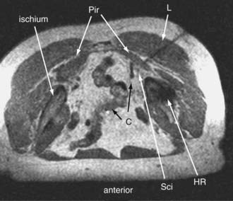
Because of the large volume of bupivacaine, procedures should be carried out in a surgicenter setting. The injection is monitored with fast (10 to 15 seconds) three-slice image sets—a working image and the two adjacent images slices. The needle advance must be maintained in the center slice of the three-slice set so that an accurate depiction of needle depth is seen. Because the piriformis muscle is often no more than 1 to 2 cm thick (Fig. 238-6), and because the bowel is often adjacent just deep to the muscle, it is necessary to reimage once or twice with each movement of the needle. Lower-accuracy alternatives to open MRI include x-ray fluoroscopy with the needle directed toward the space below the sacroiliac joint, computed tomography (CT) guidance, or electromyography with ultrasound.7


Patients obtaining complete or nearly complete and specific relief of symptoms and who fail to respond to spinal injections with similar agents can be considered to have confirmed muscle-based piriformis syndrome. If the symptoms recur within 1 week, the patients can be referred for piriformis surgery. If the symptoms recur after 2 weeks, additional injections can be carried out. Botulinum toxin injections (100 units in 6 mL preservative-free saline) can help some patients achieve longer-lasting relief from injection.


Minimal Access Surgery for Pelvic Entrapments of the Sciatic Nerve


Surgery is carried out generally on an outpatient basis using a 3-cm incision for a minimally invasive transgluteal approach with piriformis muscle resection, neuroplasty of the sciatic nerve and of the posterior femoral cutaneous nerve,8 and then placement of Seprafilm (Genzyme, Cambridge, MA) as an adhesiolytic agent. A similar approach is used for sciatic entrapments at the level of the ischial tuberosity.


For piriformis surgery, placement of the 3-cm incision is based on locating the superior medial edge of the greater trochanter of the femur with an posteroanterior hip radiograph. The patient is positioned prone on bolsters so that the knee falls below the level of the hip; this provides a relative elevation of the greater trochanter in the surgical site aiding access to the piriformis tendon.


After opening the gluteal fascia with bipolar cautery and Metz scissors, blunt finger dissection through the leaves of gluteal musculature minimizes exposure trauma and helps ensure outpatient management. Exposure is maintained with a Shadow-Line retractor system (V. Mueller)—this is an anterior cervical-type retractor that has a blade-retractor connection providing good rigidity under strong tension, but allowing for rapid replacement of blades as the depth of surgery progresses. A set of blades up to 100 mm in length (custom-made by V. Mueller) is sufficient for nearly all patients.


Safety for the sciatic nerve is ensured by carefully progressing through the muscle layers until the hard, clear prepiriformis fascia is reached with dense yellow fat behind it. The retractor blades are then reset, and the fascia is opened carefully with bipolar cautery and Metz scissors. An electrodiagnostic system with electromyographic monitoring of multiple superior gluteal, inferior gluteal, tibial nerve and peroneal nerve innervated muscles set at 0.5 to 10 mA is used to identify nerves before their exposure in the piriformis fat pad.


The sciatic nerve is partially mobilized and used, together with the greater trochanter and the sciatic notch, to identify and confirm the borders of the piriformis muscle. The femoral neck varies from nearly horizontal to nearly vertical, and the height of the greater trochanter is variable as well. In a patient with a horizontal femoral neck and large greater trochanter, the piriformis muscle can be nearly perpendicular to the sciatic nerve. By contrast, in a patient with a vertical femoral neck and short greater trochanter, the piriformis muscle is nearly parallel with the sciatic nerve.


Many patients have multipartite piriformis muscles, and in some, the superior border may appear fused with deep gluteal muscles. Some patients have an accessory piriformis muscle compressing the more proximal portion of the sciatic nerve, and this is sectioned and removed as well. Special attention and caution is required for patients in whom preoperative imaging demonstrated a split piriformis traversed by a split sciatic nerve. The slip of muscle passing between the tibial and peroneal sciatic components must be removed as well. It is always helpful to ensure by electrodiagnostic stimulation that both tibial and peroneal portions of the sciatic nerve are in view before any piriformis resection is started.


One or two silk ties are placed around the muscle so that bipolar cautery and Metz scissors can be used to fully transect the muscle in two locations with ongoing complete hemostasis. Anesthetic injection of the muscle immediately before resection can help minimize postoperative discomfort. Removal of a segment of muscle about 2 cm in length helps ensure against readhesion of the separated segments that can occur when a single cut is made. This procedure also generally severs the nerve to the piriformis muscle, resulting in subsequent atrophy of any remaining components.


Neuroplasty of the distal lumbosacral plexus, sciatic nerve, and posterior femoral cutaneous nerve is then carried out by blunt dissection generally using DeBakey pickups and a tonsil clamp. Specifically, this entails separating any abnormal fibrous covering from the nerve so that the nerve is free and fully mobile at the end of the dissection. In many cases, fibrovascular bands cross or compress the sciatic nerve and can be cut. Gentle dissection technique, liberal use of electrodiagnostic stimulation when nerve locations are in question, and meticulous, precise hemostasis with bipolar cautery before cutting any tissue help protect the neural tissues.


By swinging the retractor system, ready access can be achieved to the sciatic nerve from the top of the ischial tuberosity to well inside the sciatic notch, allowing for full mobilization of at least 12 cm of the nerve course. Decompression and muscle resection inside the pelvis through the sciatic notch is not recommended on a routine basis because of higher risk to autonomic fibers in the presacral area.


Dexamethasone, 10 mg given intravenously, can be administered at the start of the procedure. Powder-free gloves must be used to further reduce the risk for postoperative fibrosis. Only bipolar cautery is used once the gluteal fascia is reached. Meticulous and complete hemostasis is ensured before closure. Upon completion of the neuroplasty, the wound should be irrigated copiously with antibiotic irrigation maintained at body temperature in a solution warmer. Seprafilm (Genzyme) pieces should be placed in layers on all dissected nerve surfaces as an adhesiolytic agent.


Marcaine (0.5% without epinephrine) is applied to the Seprafilm and dissected nerves and is instilled in gluteal muscles along the line of approach. The gluteal fascia is then closed with 0-Vicryl sutures. Usually, no drain is placed. Patients are allowed to ambulate immediately. Patients experiencing significant muscle spasm or local pain are provided pain management in the facility overnight.

Stay updated, free articles. Join our Telegram channel

Aug 7, 2016 | Posted by in NEUROSURGERY | Comments Off on Piriformis Syndrome, Obturator Internus Syndrome, Pudendal Nerve Entrapment, and Other Pelvic Entrapments

Full access? Get Clinical Tree
Get Clinical Tree app for offline access