Fig. 32.1
The “combined endoscopic transnasal/transseptal binostril approach.” Observe the nasoseptal flap on the left side (F) and the intact right septal mucosa (SM), sphenoid rostrum (SR), inferior turbinate (IT), and middle turbinate (MT)
Then, the sphenoid rostrum is exposed and removed using a cutting burr or a Kerrison punch. This wide sphenoidotomy allows good visualization of the bony impression of the structures in the posterior wall of the sphenoid sinus, such as the optic chiasm, ICAs, sella, and clivus (Fig. 32.2).
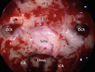

Fig. 32.2
Anatomical structures of the sphenoid sinus: prominence of the internal carotid arteries (ICA), optic canals (ON), optic–carotid recess (OCR), clivus, planum sphenoidale (PS), tuberculum sellae (TS)
32.3.2 Sellar and Parasellar Stage
32.3.2.1 Expanded Access to the Tuberculum Sellae and Planum Sphenoidale
Many pituitary macroadenomas with suprasellar extension can be removed through a standard transellar approach: the removal of sellar portions of the adenoma facilitates descent of the diaphragm sellae and suprasellar portions of the tumor into the operative field. However, in some cases, a transplanum approach may be needed [11].
The transplanum approach is the anterior-superior extension of the sellar approach. It is used when there is significant tumor extension above the sella, or when sellar access is limited by medial protrusion of the internal carotid arteries. This route provides some advantages compared with traditional craniotomies: it optimizes exposure, minimizes the risk of complications, and prevents nerve damage and excessive retraction of brain parenchyma.
To begin, the sellar floor is opened with a 4 mm diamond burr until it is eggshell-thin and then resected with a Kerrison punch. The sellar dura is exposed after bone resection with the following limits: superiorly, the superior intercavernous sinus (below the tuberculum sella); inferiorly, the inferior intercavernous sinus (superior edge of the clivus); and laterally, both carotid prominences.
Then, drilling of the thick tuberculum sellae bone and the planum sphenoidale is performed. The area between both optic canals is thinned down to eggshell thickness with a high-speed drill and then removed. This area is much wider and higher than that exposed in standard pituitary surgery (Fig. 32.3).
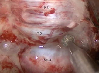

Fig. 32.3
Diamond burr used to drill out the planum sphenoidale (PS), tuberculum sellae (TS), and sellar (S) bone, exposing the dura mater of these regions during a transplanum approach
The dural opening starts at the sellar area, with a rectangular shape, after identifying the position of the ICA prominences, the cavernous sinuses (CS) and the superior and inferior intercavernous sinuses (four blue landmarks visualized through the dura) [10]. Then, the superior intercavernous sinus is coagulated with the aid of a bipolar cautery (Fig. 32.4). The dura is opened above and below the superior intercavernous sinus, exposing the sellar and suprasellar region.
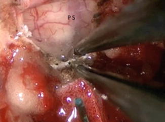

Fig. 32.4
Bipolar coagulation of the superior intercavernous sinus. Observe the dural opening of the planum sphenoidale (PS) that allows visualization of both the gyrus rectus and the arachnoid
In macroadenomas, an initial debulking is done in the inferior portion of the sellar portion of the tumor, followed by an extracapsular dissection of the lateral extension under visualization with a 45° scope and use of a curved suction tube, first identifying the angle between the arachnoid and the internal carotid artery.
In the suprasellar area, capsular dissection of the tumor under direct visualization of neural structures is favored as opposed to curettage and scraping techniques. Dissection in the plane between the tumor and normal structures facilitates complete tumor removal with maximal preservation of normal tissues, particularly the residual pituitary gland.
Suction tubes are extremely useful for keeping the surgical field clean and for aspiration of the tumor, as most pituitary adenomas have a soft consistency.
Angled endoscopes, especially 45° ones, are particularly useful for visualization each step of removal and inspection of tumor fragments near the cavernous sinus and, superiorly, in the region of the diaphragma sellae.
For tumors with superior (intraventricular) and lateral extension, intraoperative navigation using a fused CT/MRI image is useful for recognition of important structures, making the procedure safer (Figs. 32.5 and 32.6).
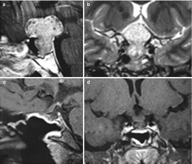
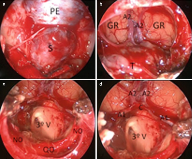

Fig. 32.5
Large pituitary adenoma with suprasellar extension. (a, c) Preoperative sagittal and coronal MRI. (b, d) Postoperative sagittal and coronal MRI

Fig. 32.6
Transplanum approach for resection of large adenoma with suprasellar extension (case of Fig. 32.5). (a) Zero-degree endoscopic view of the sella (S) and planum sphenoidale (PE) dural exposure. (b–d) Large dural opening for tumor removal through a 45° view. (T) Tumor, (A1 and A2) segments of anterior cerebral artery, (C) anterior communicating artery, (GR) gyrus rectus, (QO) optic chiasm, (NO) optic nerves, and (3°V) third ventricle
Another important technology that is part of the surgical armamentarium is intraoperative MRI (iMRI). Some authors have demonstrated that 20–30 % of intraoperative images will show additional resectable tumor, with considerable variability (5–66 %) [12].
32.3.3 Approaches to the Cavernous Sinus
Patients with pituitary tumors that invade the cavernous sinus have usually been candidates for medical treatment or radiation therapy. Surgical treatment was limited to transcranial approaches, as, under microscopic visualization, the transsphenoidal route is restricted to midline tumors. However, the transcranial route is associated with high morbidity, especially to the cranial nerves: the lateral wall of the cavernous sinus and its contents are directly in the line of surgical dissection, constituting an obstacle between the surgeon and the tumor.
Conversely, the transsphenoidal endoscopic approach can improve the lateral surgical view, making removal of tumors that invade the cavernous sinus from a medial approach—with less manipulation of cranial nerves—a feasible procedure. In this area, tumor consistency is the main prognostic factor for radical removal, as hard tumors can rarely be dissected from the carotid artery.
Stay updated, free articles. Join our Telegram channel

Full access? Get Clinical Tree


