Fig. 40.1
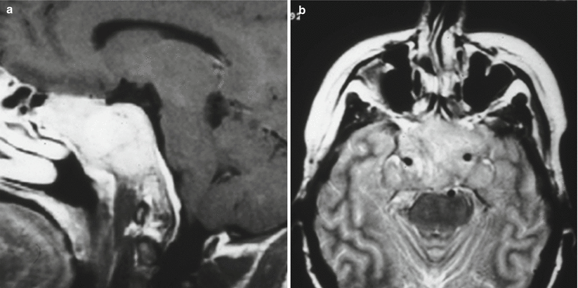
Plasmacytoma. (a) Sagittal T1-weighted precontrast image. (b) Axial T1-weighted precontrast image. (c) Axial T1-weighted gadolinium-enhanced image. There is an expansile, homogeneously enhancing mass replacing the entire clivus. The pituitary gland appears normal within the sella

Fig. 40.2
Plasmacytoma. (a) Sagittal T1-weighted gadolinium-enhanced image. (b) Coronal T1-weighted gadolinium-enhanced image. There is a large, homogeneously enhancing mass replacing the clivus and obliterating the sella. The pituitary gland is not distinctly visualized. The mass extends to bilateral cavernous sinuses

Fig. 40.3
Plasmacytoma. (a) Sagittal T1-weighted gadolinium-enhanced image. (b) Coronal T1-weighted gadolinium-enhanced image. (c) Axial T1-weighted gadolinium-enhanced image. There is a large, homogeneously enhancing mass replacing the clivus. The pituitary gland is not distinctly visualized

Fig. 40.4
Plasmacytoma. (a) Sagittal T1-weighted gadolinium-enhanced image. (b) Coronal T1-weighted gadolinium-enhanced image. (c) Axial T1-weighted gadolinium-enhanced image. There is a homogeneously enhancing mass centered in the clivus. The pituitary gland is not distinctly visualized

Fig. 40.5
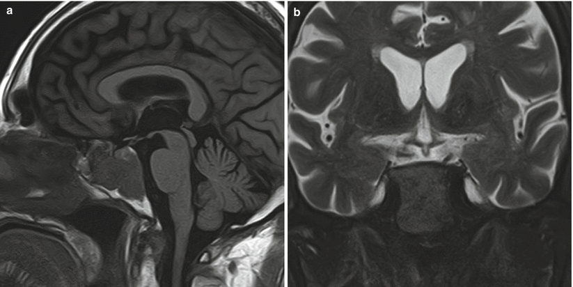
Plasmacytoma. (a) Sagittal T1-weighted image. (b) Coronal T2-weighted image. There is a large mass centered in the clivus eroding into the sphenoid sinus. The mass is distinct from the pituitary gland
40.3 Histopathology
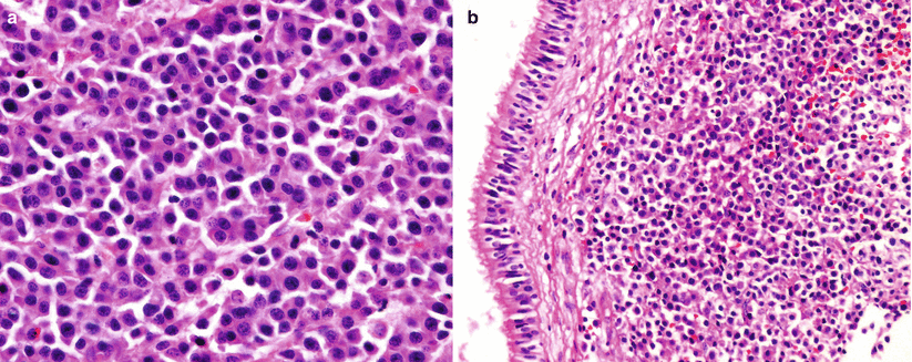
Plasmacytomas are highly cellular tumors typically consisting of a monoclonal origin of plasma B cells (Fig. 40.6).
They demonstrate positive immunoreactivity for CD138, CD43, and kappa light chain immunoglobulin. Negative immunoreactivity for chromogranin, synaptophysin, placental alkaline phosphatase (PLAP), EMA, CEA, and CAM52 is typically seen in plasmacytoma.
This diagnosis requires differentiation from lymphocytic hypophysitis (polyclonal B-cell lineage).


