• A midline posterior cervical exposure is performed.
• Care is taken not to strip the facet capsule.
• Only the medial portion of the lamina/facet junction is exposed.
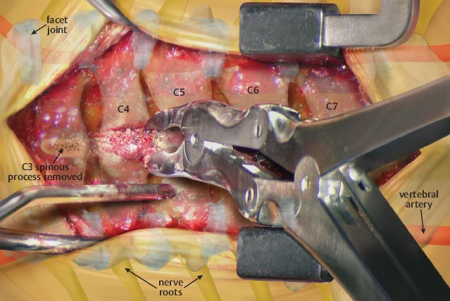
• Once exposed, the spinous processes from C3–C7 are removed.
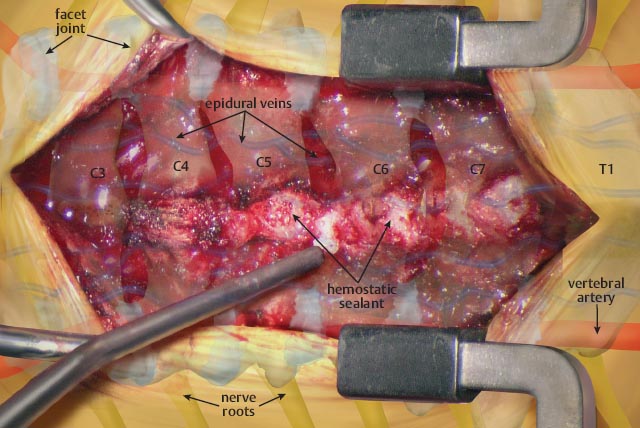
• Once the spinous processes are removed, bone wax or a hemostatic sealant is applied to control bone bleeding.
• The junction between the lamina and facet is identified.
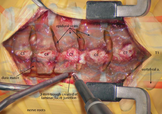
• A burr is used to create an opening of approximately 3 mm.
• This opening requires the removal of 15% of the facet joint.

Related posts:


Stay updated, free articles. Join our Telegram channel

Full access? Get Clinical Tree


