Fig. 14.1
Case 4. Surgical view of the marked line to be incised
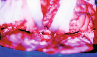
Fig. 14.2
Case 4. Surgical view under magnification showing the retraction of the frontal poles
The interhemispheric and the bilateral olfactory cisternae are opened in order to drain the CSF and to avoid further retraction. Both olfactory nerves should be dissected symmetrically without coagulation under small bleedings. Surgicel® is enough to stop the small bleedings mainly near the crivous plate of the ethmoidal bone. After that, the surgical overview could better show the image of both olfactory nerves and both optic nerves composing a groove enlarged by the tumor. The pseudocapsula of the tumor can be seen through this groove, and the dissection between the carotid artery and the pseudocapsula should be performed on both sides. Following this step, a debulking of the tumor should be done, and a piecemeal resection is recommended. The ultrasonic aspirator may be used with extreme caution. The retraction of bilateral frontal poles should never be strong enough to hurt the pia mater and cause damage to the brain parenchyma [1, 2].
14.3 Patient Population
The surgical technique described was applied in the treatment of the 11 patients, as shown in Table 14.1. Of the 11 patients, 5 were female and 6 were male, with a mean age of 37.7 years (range, 5–63 years). Histological examination revealed four adenomas (Figs. 14.3, 14.4, 14.5, 14.6, and 14.7), two craniopharyngiomas (Figs. 14.8 and 14.9), one glioblastoma, one anaplastic glioma, one meningioma, one Rathke cyst, and one schwannoma. The symptoms at admission were typical for each pathological condition, and the lesions were totally removed by using a bilateral subfrontal approach. All these patients were preoperatively evaluated with magnetic resonance and tomography. We evaluated prospectively the clinical findings at the presentation, operative treatment, and outcome of each patient.
Table 14.1
Clinical data for 11 patients with preservation of olfactory tract
Case | Gender | Age (years) | Location | Pathology |
|---|---|---|---|---|
1 | M | 24 | Left frontal fossa | Intracerebral schwarnoma |
2 | F | 54 | Sellar and parasellar | GH pituitary adenoma |
3 | F | 35 | Left frontal fossa | Anaplasic glioma |
4 | M | 30 | Sellar and parasellar | Craniopharyngioma |
5 | M | 35 | Sellar and parasellar | Non functionant pituitary adenoma |
6 | M | 36 | Tubercle sellar | Meningioma |
7 | M | 20 | Sellar and parasellar | Craniopharyngioma |
8 | M | 5 | Sellar and parasellar | Rathke cyst |
9 | F | 61 | Sellar and parasellar | Non functionant pituitary adenoma |
10 | F | 63 | Hypothalamus | GBM |
11 | F | 52 | Sellar and parasellar
Related posts: Preoperative Visualization of the Facial Nerve Using Diffusion Tensor Imaging Fibre Tracking in Patients with Large Vestibular Schwannomas Preoperative Visualization of the Facial Nerve Using Diffusion Tensor Imaging Fibre Tracking in Patients with Large Vestibular Schwannomas
 Endoscope-Assisted Microsurgery Endoscope-Assisted Microsurgery
 Restoration of Locomotion in Post-traumatic Paraplegics: The Neurosurgeon’s Personal View Restoration of Locomotion in Post-traumatic Paraplegics: The Neurosurgeon’s Personal View
 Endoscopic Transnasal Surgery for Clival Chordoma Endoscopic Transnasal Surgery for Clival Chordoma
 Diagnosis and Treatment of Adult Hydrocephalus Diagnosis and Treatment of Adult Hydrocephalus
 The Surgical Management of Trigeminal Schwannomas The Surgical Management of Trigeminal Schwannomas
Stay updated, free articles. Join our Telegram channel
Full access? Get Clinical Tree
 Get Clinical Tree app for offline access

|