Progressive Muscular Dystrophies


Progressive Muscular Dystrophies


Mathula Thangarajh

Petra Kaufmann

Louis H. Weimer

Michio Hirano

Lewis P. Rowland


CLASSIFICATION

The classification of muscular dystrophies is based on both clinical and genetic characteristics, starting with three main types: DMD, facioscapulohumeral muscular dystrophy (FSHD), and MMD. Each type differs from others in age at onset, distribution of weakness, rate of progression, presence or absence of calf hypertrophy, or high serum levels of creatine kinase (CK), and pattern of inheritance (Table 142.1).

Early investigators recognized that these three types did not include all of the common muscular dystrophies, so they included a fourth subtype of muscular dystrophy, namely, limb-girdle muscular dystrophy (LGMD). Patients with LGMD often have weakness of the shoulder and pelvic girdle muscles. In some forms of LGMD, cardiac and respiratory involvement can be prominent. The identification of several genes associated with LGMD-like phenotype has resulted in a newer classification of LGMD based on whether the disease is inherited in an autosomal dominant or recessive pattern.

Progressive muscular dystrophies result from diverse defects in muscle proteins associated with the extracellular matrix (collagen type VI, merosin); cell membrane and associated proteins (dystrophin, sarcoglycans, caveolin-3, dysferlin, integrins); cellular enzymes (calpain-3), organelle, or sarcomere function (telethonin, myotilin, titin); and nuclear envelope (lamins, emerin) (Fig. 142.1).


X-LINKED MUSCULAR DYSTROPHIES


PREVALENCE AND INCIDENCE

The incidence of DMD is about 1 in 3,500 male births, with no geographic or ethnic variation. Approximately one-third of the
cases are caused by de novo mutations; the others are more clearly familial. Because the life span of patients with DMD is shortened, the prevalence is less—about 1 in 18,000 males. Becker muscular dystrophy (BMD) is much less common, with a frequency of about 1 in 20,000.


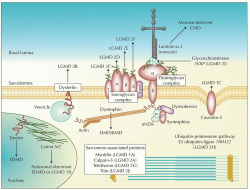
FIGURE 142.1 Skeletal muscle proteins and muscular dystrophies. The dystrophin-glycoprotein complex (DGC) comprises dystrophin, the dystroglycans (α, β), the sarcoglycans (α, β, γ, δ), sarcospan, the syntrophins (α, β1), and dystrobrevin (α). The complex stabilizes the sarcolemma and protects surface membranes in muscle contraction. Nitric oxide synthetase (nNOS) interacts with the syntrophin complex and laminin-α 2, one of many extracellular ligands of α-dystroglycan. Disease-causing mutations are cited in the text and Table 142.3. LGMD, limb-girdle muscular dystrophy; CMD, congenital muscular dystrophy; DMD, Duchenne muscular dystrophy; BMD, Becker muscular dystrophy; EDMD, Emery-Dreifuss muscular dystrophy. (From Mathews KD, Moore SA. Limb-girdle muscular dystrophy. Curr Neurol Neurosci Rep. 2003;3:78-85, with permission.)


Duchenne and Becker Muscular Dystrophies


DUCHENNE MUSCULAR DYSTROPHY

The condition may become evident in infancy if serum enzymes are coincidentally measured, for example, for an incidental respiratory infection. A positive family history may also alert an astute physician to the early recognition of motor delay. Authorities often state that symptoms do not begin until age 3 to 5 years but that view may be a measure of the crudeness of muscle evaluation in infants. Infants with DMD score less on scales that evaluate gross motor function. Many boys have hypertrophied calf muscles. Walking is often delayed, and the boys probably never run normally; toe walking and waddling gait are evident. Then, the condition progresses to overt difficulty in walking, climbing stairs, and rising from chairs. An exaggerated lordosis is assumed to maintain balance. The boys tend to fall easily if jostled and then they have difficulty rising from the ground. In doing so, they use a characteristic maneuver called the Gowers sign (Fig. 142.2). They roll over to kneel, push down on the ground with extended forearms to raise the rump and straighten the legs, then move the hands to the knees and push up to a standing position. The process has been called climbing up himself. It is also seen in other conditions that include proximal limb and trunk weakness, such as spinal muscular atrophy. At this stage, the knee jerks may be lost, whereas ankle jerks are still present; this discrepancy is a measure of the proximal accentuation of weakness.

As the disease progresses, the arms and hands are affected. Slight facial weakness may be seen, but speech, swallowing, and
ocular movements are spared. Iliotibial contractures limit hip extension; heel cord contractures are partly responsible for toe walking. Loss of ambulation occurs between ages 9 and 12 years or later when treated with steroids. Boys then become wheelchairbound. Scoliosis may then become serious and may compromise limb and respiratory function. Elbow and knee contractures contribute to disability. Respiratory muscle weakness causes progressive decline in lung capacity beginning at about age 8 years. Nocturnal hypoventilation may occur and cause morning headaches and daytime fatigue if untreated. By about age 20 years, respiration is severely compromised that respiratory support is needed. Life expectancy in DMD has improved greatly in the last three decades, probably because of better coordinated medical care and home nocturnal ventilation. Some attribute the improvement to prednisone therapy.


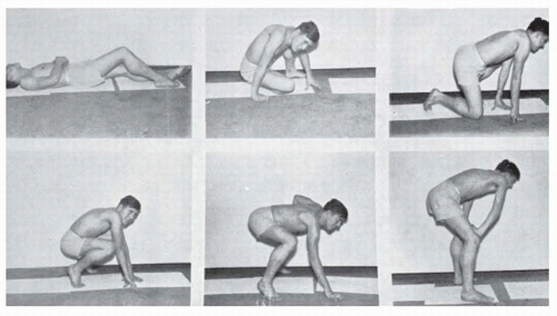
FIGURE 142.2 Gowers sign in a patient with DMD or BMD. Postures assumed in attempting to rise from the supine position.

The heart is usually spared clinically until late in the disease when congestive heart failure (CHF) and arrhythmias may develop. The ECG is abnormal in most patients, with increased QRS amplitude in lead V1 and deep, narrow Q waves in left precordial leads. Echocardiography shows progressively diminished contractility. Signs of cardiomyopathy may not be apparent because of the inability to exercise but may supervene in a few cases. The gastrointestinal system is usually spared, but intestinal hypomotility and acute gastric dilatation are uncommon complications probably related to dystrophin deficiency in smooth muscle. Osteoporosis begins in the ambulatory years, is more severe in the legs, and may contribute to fractures. Intellectual impairment with predominantly verbal difficulties is seen in one-third of boys with DMD, and the average IQ is shifted approximately 1 standard deviation below normal. Therefore, some boys with DMD have average or below average intelligence.

Anesthetic catastrophes may occur with hyperkalemia, extremely high CK levels (even more than 20 times the normal maximum), acidosis, cardiac failure, hyperthermia, and rigidity. The risk can be decreased by avoiding inhaled general anesthetics and depolarizing muscle relaxants, especially succinylcholine. This vulnerability can be considered a form of malignant hyperthermia.


BECKER MUSCULAR DYSTROPHY

This condition is defined by milder skeletal muscle weakness compared to DMD. Calf hypertrophy is present, weakness is greatest proximally, and serum CK levels are high. EMG is myopathic and muscle biopsy reveals some staining of dystrophin. The two clinical features that distinguish it from DMD are differences in age at onset (typically after age 10 years) and rate of progression (still walking after age 16 years, often later). Cognitive impairment is less common than in DMD. Cardiomyopathy usually develops later but may overshadow limb weakness. Cardiac complications contribute to 50% of BMD deaths compared with 20% with DMD. However, some patients have severe cardiomyopathy and mild muscle weakness. Another allelic disorder, X-linked dilated cardiomyopathy, demonstrates little to no skeletal muscle weakness.


INTERMEDIATE PHENOTYPE

Some patients show an intermediate phenotype that can be considered either severe BMD or mild DMD. They remain ambulatory after age 12 years but use wheelchairs before age 16 years. They have preserved antigravity strength in the neck flexors longer than typical DMD patients.


MANIFESTING CARRIERS

DMD is an X-linked recessive condition. Female mutation carriers are usually asymptomatic, but “manifesting carriers” can have limb weakness, calf hypertrophy, cardiomyopathy, or high serum CK levels.


MOLECULAR GENETICS

The dystrophin gene is involved in both Duchenne and Becker dystrophies, which are allelic diseases. Dystrophin is a cytoskeletal protein located at the plasma membrane. Brain and other organs contain slightly different isoforms. In muscle, dystrophin is associated with membrane glycoproteins that link the cytoskeleton with membrane proteins and then to laminin and the extracellular matrix (see Fig. 142.1). The protein probably plays an essential role in maintaining muscle fiber integrity. If dystrophin is absent, as in Duchenne dystrophy, or is abnormal, as in Becker dystrophy, the sarcolemma becomes unstable in contraction and relaxation, and the damage results in excessive calcium influx, thereby leading to muscle cell necrosis. If specific glycoproteins are abnormal or missing, the same problems arise, as in some LGMDs (described later in the chapter).

These findings have been put to practical use in the diagnosis of Duchenne and Becker dystrophies and in genetic counseling. DNA analysis demonstrates a deletion or duplication at Xp21 in about 65% of DMD and 85% of BMD patients. Patients with DMD typically have “frameshift” mutations that alter the DNA reading frame and consequently change amino acids downstream of the mutation, whereas individuals with BMD have milder “in-frame” rearrangements that maintain the sequence of amino acids flanking the mutation. Point mutations or small rearrangements account for the remainder and can be detected by gene sequencing. The presence of a deletion or point mutation in a patient with compatible clinical findings is diagnostic. Carriers can be similarly identified, and the test can also be used for prenatal diagnosis (Fig. 142.3).

The initial evaluation of a boy suspected to have dystrophinopathy includes a serum CK followed by genetic screening for dystrophin mutations performed on peripheral blood leukocytes. Mutations that interfere with the expression of dystrophin cause DMD and those that leads to abnormal quality or quantity of dystrophin cause BMD. Muscle biopsy is sometimes performed in sporadic cases of unclear phenotype, even when a deletion has been identified, because immunocytochemistry for dystrophin and Western blot for quantitative dystrophin analysis can confirm and refine the diagnosis in these questionable cases. If no deletion is found and if dystrophin sequencing is not available, muscle biopsy is needed.


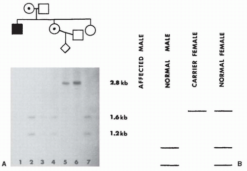
FIGURE 142.3 Prenatal diagnosis in DMD. A: Autoradiograph of Southern blot of 1% agarose gel with DNA samples from each individual digested with the restriction enzyme Xmnl and probed with pERT87-15. The affected male is deleted (no signal). His sister who was pregnant has a deleted X and an X chromosome with the 1.6/1.2 allele. Her husband’s X chromosome contains the 2.8 allele. The fetus contains the husband’s X and the deleted X. B: Diagram of the four possible outcomes of the prenatal diagnosis. The fetus is a carrier female. (Courtesy of A. D. Roses.)

In Duchenne dystrophy, dystrophin staining is absent by both immunocytochemistry and Western blot. In Becker dystrophy, immunocytochemistry shows an interrupted pattern of staining on the surface membrane, and the Western blot shows decreased dystrophin levels or abnormal protein size. Carriers show a mosaic pattern; some fibers contain normal dystrophin staining and others show no dystrophin staining. The most widely used animal model of DMD is the mdx-23 mouse, which has a spontaneous mutation in the exon 23 of the murine dystrophin gene.


OTHER XP21 MYOPATHIES


Dystrophinopathies

A deletion in a neighboring gene can extend into the dystrophin gene and produce myopathy. The resulting syndrome may be dominated by other neighboring gene changes, including congenital adrenal insufficiency or glycerol kinase deficiency, as well as a myopathy, which may range from mild to severe. These syndromes are customarily called Becker variants if appropriate dystrophin abnormalities are found. That nomenclature may be confusing, however, because the original definition of Becker dystrophy was based on clinical criteria, and these syndromes are clinically different. Among them are myopathies that affect girls or women, not only manifesting carriers but also girls with Turner syndrome or balanced translocations that involve Xp21, as well as syndromes of atypical distributions of weakness, such as distal myopathy or quadriceps myopathy. Some syndromes lack limb weakness but are manifested by other symptoms, such as recurrent myoglobinuria or X-linked cramps without weakness. Each of these atypical syndromes warrants studies of dystrophin.


Nondystrophin-Related Xp21 Myopathies

McLeod syndrome (MIM 300842) is a multisystemic disorder first discovered in blood banks because the donors lacked a red cell antigen, the Kell antigen. These individuals were found to have abnormally shaped red blood cells (acanthocytes), and serum CK values were often 29 times normal or even higher. In addition, some had myopathic limb weakness, and the condition mapped to Xp21. Dystrophin is normal in these patients. Deletions in the XK gene cause McLeod syndrome and a chromosomal microarray analysis may be helpful. It is also described in Chapter 134 (acanthocytes).


EMERY-DREIFUSS MUSCULAR DYSTROPHY

Emery-Dreifuss muscular dystrophy (EDMD) is a muscular dystrophy with several distinct manifestations:


  • The characteristic humeroperoneal distribution of weakness is unusual. That is, the biceps and triceps are affected rather than shoulder girdle muscles, and distal muscles are affected more than proximal muscles in the legs. The limb weakness may be mild or severe.


  • Contractures are disproportionately severe and are evident before much weakness is noted. The contractures affect the elbows, knees, ankles, fingers, and spine. A rigid spine develops, and neck flexion is limited.


  • Heart block, atrial paralysis, and atrial fibrillation are found in 95% of patients by age 30 years and lead to placement of a pacemaker. Dilated cardiomyopathy occurs in about 35% of all cases.

EDMD has been associated with six genes. It is characteristically an X-linked recessive disease (MIM 310300) and is linked to mutations in the EMD or emerin gene on Xq28. Emerin is localized to the nuclear membrane in muscle and other organs. Alternatively, the phenotype is inherited in an autosomal dominant (MIM 181350) or recessive pattern (MIM 604929) linked to the LMNA gene on 1q21, encoding lamin A/C. Lamin A/C is located in the inner nuclear lamina and has also been linked to several allelic disorders, as discussed later in the chapter. Mutations in FHL1 encoding four-and-a-half LIM protein 1 also cause X-linked EDMD. Autosomal dominant EDMD can also be caused by the following genes: SYNE1, SYNE2, and TMEM43.

Symptoms of EDMD often start before age 5 years with difficulty walking. Examination typically shows symmetric contractures and weakness without pseudohypertrophy. Tendon reflexes are hypoactive or absent, and there may be mild facial weakness. Serum CK activity can be mildly elevated to levels less than 10 times normal. The diagnosis is usually confirmed by DNA testing. Absence of emerin can be demonstrated by immunocytochemical study of a muscle biopsy. Emerin staining is absent not only in muscle nuclei but also in circulating white blood cells (WBCs) and skin. As a result, skin biopsy, leukocytes, or inner cheek swabs for exfoliated mucosal cells can be examined diagnostically instead of muscle biopsy. However, DNA or gene product analysis is preferred to establish accurate diagnosis. Cardiac surveillance (including 24-hour ECG and echocardiography) is crucial.

EDMD must be distinguished from other conditions. The rigid spine syndrome includes vertebral and limb contractures but not cardiomyopathy or muscle wasting. Because the cardiac abnormality may not be evident in childhood, some patients with a rigid spine might be expected to have EDMD. Collagen type VI-related disorders (Bethlem myopathy/Ullrich CMD—MIM 158810/254090) include contractures and myopathy but not cardiomyopathy, as discussed later in the chapter.

Management is symptomatic and may include physical therapy and surgical tendon release for contractures. Pacemaker implantation and treatment of cardiomyopathy is often crucial. Female carriers are at risk of arrhythmias and should have regular cardiac evaluations.


FACIOSCAPULOHUMERAL MUSCULAR DYSTROPHY


MOLECULAR GENETICS

The disease is autosomal dominant with nearly complete penetrance. Almost all FSHD patients have deletion of the subtelomeric array of 3.3-kilobase repeat elements, termed D4Z4, on chromosome 4q35. Normal individuals have 11 to 150 repeats; FSHD patients usually have fewer than 11. The deletion results in inappropriate expression of the double homeobox-containing gene DUX4 in muscle cells. This common form is referred to as FSHD1. About 5% of FSHD patients have chromatin relaxation at D4Z4 without D4Z4 contraction and are due to mutations in the chromatin modifier gene SMCHD1. This rare form is referred to as FSHD2.


CLINICAL MANIFESTATIONS

The following features are characteristics of FSHD:


  • Facial weakness is evident not only by limitation of lip movements but also in the slightly everted lips and wide palpebral fissures. Patients may state that they have never been able to whistle or blow up a balloon. Some are said by relatives to sleep with eyes open.


  • Scapular winging is prominent. Protrusion of the scapulae is more evident when the patient tries to push against a wall with elbows extended and hands at shoulder level. The winging becomes more evident when the patient tries to raise the arms laterally. The patient cannot raise the arms to shoulder level even though there is no weakness of the deltoids on manual testing. This limitation is caused by inadequate fixation of the scapulae to the chest wall.


  • The shoulder girdle has a characteristic appearance. Viewed from the front, the clavicles seem to sag and the tips of the scapulae project above the supraclavicular fossa. This abnormality becomes more marked when the subject tries to raise the arms laterally to shoulder level. Smallness of the pectoral muscles affects the anterior axillary fold, which is ordinarily diagonal but assumes a vertical position. Abdominal muscle weakness may cause abdominal protrusion and exaggerated lumbar lordosis. The lower abdominal muscles are weaker than the upper causing a positive “Beevor” sign with the navel moving toward the head upon neck flexion in a supine position. The limb muscle weakness is often asymmetric.


  • Leg weakness may affect proximal muscles or, more often, the tibialis anterior and peroneals, resulting in footdrop.

In family studies, asymptomatic individuals can be identified by mild versions of these signs. Within a single family, the condition may vary markedly. Progression is slow, however, and the condition may not shorten longevity. About 20% of patients eventually use a wheelchair. Respiratory insufficiency is rare. Men may be more severely affected than women.


ASSOCIATED DISORDERS

Mild sensorineural hearing loss and vascular retinopathy as well as oropharyngeal symptoms, Coats disease (exudative telangiectasia of the retina), and, possibly, mental retardation are occasionally seen in children with FSHD. However, these findings are not consistent, and it is not known how they relate to the genetic abnormality. Cardiac arrhythmias are rarely seen in FSHD.


LABORATORY STUDIES

In most cases, the diagnosis can be confirmed by leukocyte DNA testing for a contraction of the subtelomeric repeat array on 4q35. Presymptomatic diagnosis is possible in families with more than one affected member, but testing should be done only with appropriate counseling. EMG and muscle biopsy, by definition, should show a myopathic pattern. The histologic changes are mild and nonspecific. Inflammatory cells are found in some patients, raising the question of polymyositis, but immunosuppressive therapy has always failed in these patients. DNA diagnosis resolves this possible confusion and may obviate muscle biopsy. Some patients with typical manifestations do not show the characteristic mutation, which may imply locus heterogeneity (mutation at some other locus) or could exceed the limits of sensitivity of current tests for the mutation.


Jul 27, 2016 | Posted by in NEUROLOGY | Comments Off on Progressive Muscular Dystrophies

Full access? Get Clinical Tree

Get Clinical Tree app for offline access