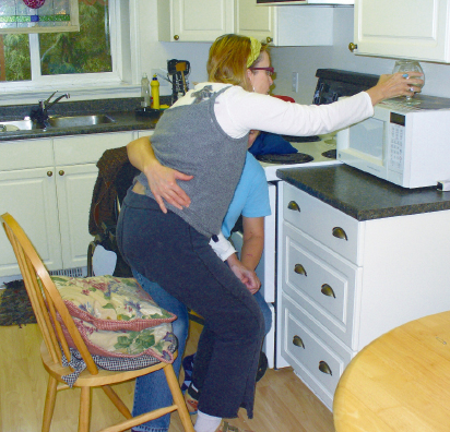
Case Report A3 Identifying and Addressing the Impairments in an Individual Who Demonstrates Contraversive Pushing Behavior Pusher syndrome was first clinically described by Davies1 in her book Steps to Follow in 1985. Contraversive pushing, also referred to as pusher syndrome, pushing behavior, or ipsilateral pushing, is defined when an individual who has had a stroke actively pushes away from the nonparetic side, leading to a loss of postural balance and falling toward the paralyzed side.2 Clinically, these individuals strongly resist passive correction to midline in both the frontal and the sagittal planes, especially if this correction is done abruptly or without any functional or task context. Most authors cite the incidence of pushing behavior as ~10% of the acute stroke population (5.3% of the entire stroke population and 10% of the stroke population who receive rehabilitation).3,4 However, other studies, including Danells et al,5 in 2004 (63% of the stroke population), and Lafosse et al,6 in 2005 (40–50% of the stroke population), report the incidence to be higher. Initially, the pushing behavior was believed to occur almost exclusively in individuals who had right brain lesions.1 However, it is now widely accepted that pushing behavior may be present in individuals with lesions in either hemisphere.2,3,7 Karnath and colleagues2,7,8,9,10,11 have been conducting extensive research with this population with various collaborators to determine the origin of pushing and to develop management strategies and techniques for its treatment. This research has determined that pushing is associated with unilateral lesions of the left or right posterolateral thalamus and leads to an altered perception of the body’s orientation in relation to gravity.2,7,8,9,10,11 In the Karnath et al2 study of 2000, both the visual and the vestibular systems were determined to be intact in these individuals. In addition, these researchers determined that, on average, these individuals perceive their upright middle at ~18° to the ipsilesional, nonparetic side. This finding led the researchers to describe a second pathway for organizing movement in gravity (separate from the one for perception of the visual world). They call this second pathway the graviceptive system. Spatial neglect and other neuropsychological impairments were traditionally suspected to be the causes of this pushing behavior, but investigations have ruled these causes out.3,9,11 There are, however, strong correlations between pushing behavior and spatial neglect after right hemisphere lesions (incidences from 100%,2,12 80%,13 67%,10 62%,5 to 40%3) and between pushing behavior and aphasia after left hemisphere lesions (incidences from 100%,9 80%,6,14 60%,10 to 47.1%3). These strong correlations (spatial neglect with right hemisphere lesions and aphasia with left hemisphere lesions) occur because the relevant brain structures associated with these functions lie in close proximity to each other.7 There is some debate among researchers about which group of individuals should be classified as pushers based on the amount of motor recovery present on the more involved side of the body (limbs and trunk).5,9,10,15 Some believe that there are two different categories; if there is severe involvement, including essentially no motor recovery on the more involved side of the body, these individuals are categorized as pushers.3,5,9,10 If there is relatively good motor recovery on the more involved side, these individuals are not categorized as pushers, despite their very active pushing away from the less involved side with active resistance to correction to vertical midline. Danells et al5 and others2,3 suggest that the recovery of pushing is not strongly associated with the recovery of motor control. Pushing is often completely resolved by 3 months (71% of subjects in the study by Danells et al5 in 2004) or by 6 months (100% of subjects in Karnath et al9,10 2002 and 2005, respectively, and the work of Pedersen et al3 in 1996), yet profound motor deficits/paresis in all these groups still existed. In 2007, Santos-Pontelli et al15 published a case report that examined pushing behavior in an individual poststroke but with only minimal paresis. Their results showed that, as the pushing behavior decreased, the functional outcome measures increased (using the Barth-el Index), yet the paresis level did not statistically change. Thus it appears that the resolution of the pushing behavior is associated with improved functional outcomes but not with resolution of paresis or whether the paresis is mild or severe. From a motor stabilizing perspective, clients who demonstrate pushing behavior do the opposite of what clients with stroke who don’t push do. Pederson et al3 in the Copenhagen Stroke Study concluded that “ipsilateral pushing did not affect functional outcome, but slowed the process of recovery considerably.” In the same report, the subjects required ~3.6 weeks longer to reach the same functional outcomes as individuals without ipsilateral pushing behavior.3 In 2004, Danells et al5 found the length of stay (LOS) to be 4.57 weeks longer to reach the same functional levels for individuals who push compared with individuals who do not push. Currently, with shorter LOSs in most Western countries and the pressure in these health care systems to move people along the continuum of care as fast as possible, it could be that individuals who push poststroke may no longer be given the opportunity to achieve the same functional outcomes given that they require more recovery and practice time. Also, as already discussed, given that their paresis is typically more severe, these individuals require more manual effort to mobilize and/or require the use of mechanical lifts for transfers and in therapy. Thus these individuals may be more likely to be directed to long-term care facilities from acute care without being considered for other programs, such as slow stream rehabilitation. It is also possible that, in acute care, they are not stood, transferred, and generally mobilized in therapy and on the medical units as often as individuals who do not demonstrate pushing behavior. Understanding and applying strategies to help those who demonstrate pushing behavior integrate and resolve the pushing as early as possible in the recovery phase may enable clinicians to assist clients with shortening the period of pushing and may thereby facilitate earlier return to the same levels of functional independence as clients who do not push poststroke. Pushing behavior and its management are not consistently described and taught in therapy schools. Novice therapists, if they have limited or no didactic knowledge or practical strategies for evaluating and working with these individuals, typically have to rely on what more experienced colleagues teach them when they first encounter an individual who demonstrates pushing behavior. The following case report demonstrates the clinical application of the Neuro-Developmental Treatment (NDT) Practice Model to severe contraversive pushing behavior. It also shows that determining the underlying cluster of system impairments contributing to the pushing behavior plays a critical role in establishing functional outcomes and developing intervention strategies for this individual who pushes. A broader discussion of contraversive pushing behavior in individuals poststroke can be found in Chapter 11 on stroke. Carol R. is a 62-year-old woman who suffered a large right ischemic frontoparietal infarct on November 17, 2008, with resultant left hemiparesis. Her comorbidities are hypothyroidism (well controlled by medication) and a past stroke (~20–25 years previously). Carol and her husband report that she fully recovered from this past stroke and had no residual deficits. Carol experienced several complications while in the hospital. Her hospital course included a pulmonary embolus, pneumonia, and insertion of a percutaneous enterogastric tube (PEG) on February 4, 2009, for nutritional purposes. This replaced the nasogastric tube she had in situ since her admission. She was moved to the inpatient rehabilitation unit ~2 weeks after her stroke and remained there until her discharge home on March 4, 2009. The stroke team encouraged Carol’s husband, Bob, to pursue complex care placement instead of taking her home—Bob refused. On discharge, referrals to home and community care occupational therapy and physical therapy and to home support (twice a day for personal care) were made. There were no options offered for outpatient therapy; the team felt Carol’s prognosis for further recovery was minimal. Carol’s swallowing abilities improved gradually once home such that the PEG could be removed by approximately May 2009 (2 months postdischarge from the hospital). Carol began private physical therapy in April 2009. Carol (and Bob’s) goals are to “fully regain her function so that she can sew, walk/hike, and work out at the gym, travel, and drive.” She wants to be “normal again and get her life back.” Carol was healthy and active prior to this stroke. She is married to Bob and has three grown children and two grandchildren. Their children and grandchildren live in different provinces. Her children provide emotional support but are not able to provide or assist in any physical care for Carol. Carol’s husband, Bob, is supportive and determined to keep her at home. He is willing to assist in any way that helps her improve, including encouraging her to do as much as she can on her own. Carol and Bob live in a one-story home in a small town in British Columbia. There are two steps to get into the home from outside. The house is all on one level indoors and is in a community with paved sidewalks and large green spaces. Their yard is full of fruit trees and flowers, and they have a small herb garden. At the time of her stroke, Carol had retired from nursing. She owned a one-woman sewing business for children’s clothing and rag dolls that she ran from her home. Carol had begun this sewing business while still working as a nurse. She sold these items on consignment at local retail stores and at craft sales and fairs. Her hobbies included reading, swimming, working out at the gym, running and hiking, traveling to see her children and grandchildren, and yard work. She shared the cooking responsibilities with her husband but not cleaning (she happily left this to her husband). Poststroke, Carol had no cognitive deficits and was able to participate in her partnership with her husband in decisions about the house, family, finances, and so forth. She is chatty and social. Presently, she is not able to participate in her occupational and social activities, such as working out at the gym, walking/hiking with her friends/spouse, performing any aspect of her sewing business, driving to the clothing stores and craft fairs where she sells her products, assisting with the gardening and lawn care, assisting with the grocery shopping, or traveling alone via plane to visit her children and grandchildren. The following general description of Carol’s activity and activity limitations give an overall impression of Carol’s functional status at the time of the initial evaluation. Carol is totally physically dependent on one person for all home and community tasks except for the following: • Eating at the kitchen table with her right hand once set up and wearing a bib (in a supportive wheelchair). • Wiping drool from her mouth with a cloth with her right hand when propped up in bed or in her supportive wheelchair. • Watching TV and reading books while propped up in bed or in her supportive wheelchair. Observational posture and movement analysis was used to establish and document Carol’s baseline functional performance level in functional tasks she performed during the initial evaluation. These same activities were reevaluated and measured periodically, as will be shown later in this case report. 1. Transfers (wheelchair ↔ bed, lying ↔ sitting, wheel-chair ↔ commode, wheelchair ↔ car) Note: Home and community care home support staff (the care aides) were not allowed to transfer Carol because her transfers were deemed to be too heavy for them to perform. Based on their WorkSafe policies, she would be classified as needing a mechanical lift if the care aides were to perform the transfers. Fig. A3.1 Carol’s sitting posture on initial examination and evaluation. 2. Sit to stand for pulling pants up. 3. Sit to lying to get into bed to right side. 4. Indoor mobility. Note: Carol reported that she stood only once during her inpatient rehabilitation stay. 5. Toileting. 6. Applying makeup. 7. Other performance. Bob built a ramp to accommodate Carol’s wheelchair getting in and out of the house. They installed a bedrail under the mattress on the right side (Carol’s side) and purchased a lightweight wheelchair with a Quadtro Select Cushion (ROHO, Inc.) and a Personal Backrest (Invacare Corp.). Carol and Bob also purchased a commode chair for toileting because both bathrooms were too small for her wheelchair and a caregiver and a safe transfer. • Inappropriate overrecruitment of right lateral trunk flexors and limb extensor muscles (UE > lower extremity [LE]), specifically, concentric activity of these muscles. Does not accept weight onto the right limbs, with eccentric > isometric muscle contractions. (This is an impairment specific to individuals demonstrating contraversive pushing.) • Decreased perceptual awareness of midline frontal plane > sagittal plane. (This is an impairment specific to individuals demonstrating contraversive pushing behavior.) • Tightness in the following: Clinical Scale for Contraversive Pushing Score = 5.5 (See Chapter 11 on stroke for more details on the Scale for Contraversive Pushing.2) Individuals poststroke who demonstrate pushing behavior have system impairments similar to those of individuals poststroke that do not push. These include impairments in the neuromuscular system, such as the inability to initiate or sustain activity and weakness in the muscles on the more involved side of the body (trunk and limbs); the musculoskeletal system, such as shortening of muscles and other soft tissues, typically on the more involved side of the body (trunk and limbs); the sensory/perceptual system, such as loss of or diminished awareness of proprioception, light touch, and mid-line orientation; and the cardiovascular system, such as decreased endurance. However, as was discussed in Chapter 11, individuals who demonstrate contraversive pushing poststroke also have a subset cluster of impairments specifically related to the pushing behavior. In Carol’s case, the impairments specifically related to her pushing behavior are the inappropriate overrecruitment of activity in the right lateral trunk and limb extensor muscles (UE > LE); specifically, concentric activity of these muscles (she does not accept weight onto her right side (trunk and limbs) with eccentric greater than isometric muscle contractions, and her decreased perceptual awareness of mid-line in the frontal greater than sagittal planes. Private home-based physical therapy began April 10, 2009. She was seen approximately twice a week for therapy sessions between this time and the fall of 2010. After this time, the frequency was approximately once a week until the spring of 2011. This case report covers the time up to the end of May 2010. Carol’s therapy continues (after the spring of 2011) with bursts of sessions on an ongoing basis. She continues to make steady, slow gains. Included at the end of this case report is an addendum with Carol’s progress after the spring of 2010. Individuals who demonstrate pushing behavior have system impairments similar to those of individuals who demonstrate more typical poststroke impairments, plus a subset of impairments specifically related to the pushing. This subpopulation of individuals poststroke who demonstrate contraversive pushing behavior requires a further collection of intervention strategies and ideas to address their specific collection of impairments. A more detailed collection of these strategies can be found in Chapter 11. Examples of the intervention activities used with Carol during her therapy sessions follow. Throughout the therapy sessions, the therapist strived to keep Carol’s left hand/arm in closed chain setups where possible; this occurred more during the mid to later time frame when the need for trunk and leg cues and support decreased and the therapist had a free hand occasionally. Therapy sessions occurred in her kitchen, sewing room, outside in the yard and garden when the weather permitted, in her bedroom, in her two bathrooms, and at the community pool. The activities were varied to keep her and the therapist’s interest. Her husband was present during all the sessions (other than when in the pool—he used this time for respite). Bob both helped as needed and also learned from what was done during the therapy sessions so that he could carry over the practice strategies between therapy sessions. Fig. A3.2 shows a kitchen activity working in the postures of liftoff and squat. The intervention activities are divided into three phases; early-, mid-, and late-phase time frames to show some of the intervention progressions made as Carol’s pushing tendencies decreased and as her strength and function improved.
A3.1 Introduction
A3.2 Case Description
A3.2.1 Personal Goals
A3.2.2 Examination and Evaluation
Social, Environmental, and Contextual Factors
Participation and Participation Restrictions
Activity and Activity Limitations
Baseline Functional Performance Measures as of April 2009
 Required maximum assist of one person.
 Helper (always her husband Bob).
 Performed all wheelchair setup including brakes, footrests, and armrests and positioning of her feet.
 Braced her feet and used a transfer belt around her midtrunk to pull her to standing and pivot from seating surface to seating surface.
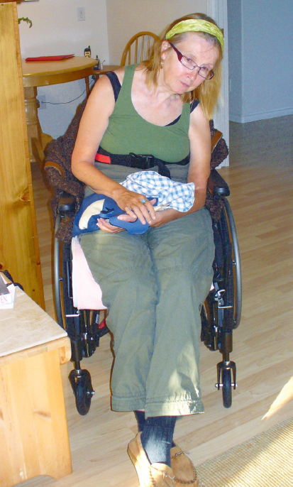
 Carol (Fig. A3.1).
 Pushed backward and to the left with right arm > leg during the transfers.
 Right foot tended to reposition constantly to the right and forward of her center of mass (COM).
 Trunk remained in marked thoracic flexion with shortening and posterior rotation of the rib cage on the left side, forward head posture with left neck side flexion.
 Left upper extremity (UE) hung at her side with palpable three fingerwidth glenohumeral subluxation.
 Setup, alignment, and assist are as described in list item 1.
 Once in standing, Carol grabbed onto a clothes dresser or braced her self against the wall so Bob could use one of his hands to pull Carol’s pants up.
 Alignment as described in list item 1.
 Fell backward and to the left if left unsupported in sitting.
 Attempted to assist with right arm on bed for sitting to side lying but pushed away and back from the right side and thus was only successful ~20% of the time, requiring constant verbal and tactile cueing to use this arm in a helpful way.
 Helper lifted full weight of both legs from floor into bed and repositioned legs for comfort.
 Helper assisted with upper body positioning once in supine.
 Nonambulatory.
 Occasionally wheeled wheelchair with right hand and right foot/leg short distances only; ~2 to 3 m on smooth, level surfaces in a forward or backward direction.
 Unable to make turns or negotiate carpets or ramps in wheelchair.
 Used bedside commode chair only for toileting (not going into bathroom at all) with assistance of husband and caregivers.
 Assisted as in list item 1.
 Required supervision of one for balance while sitting on commode.
 Wipes her face with a cloth and applies the makeup with her right hand when sitting in a supportive wheelchair in front of a mirror.
 Trunk alignment is as described in list item 1.
 Once her feeding tube was removed, Carol began once-weekly tub baths at a nearby assisted living facility. A mechanical lift was used to transfer her in and out of the tub. The home support workers bathed her.
 Carol used a cloth to wipe drool from her mouth with her right hand approximately every 2 to 3 minutes while sitting.
Equipment
Carol’s Most Significant System Impairments
Neuromuscular
 Inability to sustain activity in spine extensor muscles (thoracic >> lumbar and left [L] >> right [R]).
 Weakness in left hip abductors, extensors, and quadriceps muscles.
 Weakness in left lateral trunk muscles.
 Inability to initiate activity throughout left UE, including all of the scapular muscles.
 Inability to initiate activity in all left LE muscles other than hip abductors, extensors, and quadriceps as described.
Sensory/Perceptual
Musculoskeletal
 All neck muscles for side flexion, rotation and long pivot extension (L > R).
 PF muscles; L >> R, and soleus > gastrocnemius muscles.
 Left lateral trunk muscles (including rotators).
 Left glenohumeral (GH) internal rotators and adductors.
 Left metacarpophalangeal (MCP) and proximal interphalangeal (PIP) extensors (fourth digit especially).
Cardiovascular
General Cardiovascular Deconditioning
A3.2.3 Intervention
Related posts:
 The Practice of Speech-Language Pathology from a Neuro-Developmental Treatment Perspective
 Report B5 Enhancing Functional Independence in a 10-Year-Old Boy with Cerebral Palsy, Spastic Quadriparesis
 Motor Control
 Report B3 Development of an Intervention Plan of Care for a Young Child with Hemiplegia
 Report B1 Multidisciplinary Examination and Intervention Planning for Identical Twin Infants with Extreme Prematurity
 Evaluation and Developing the Plan of Care
![]()
Stay updated, free articles. Join our Telegram channel

Full access? Get Clinical Tree


