14 Rickettsial and mycoplasma infections RICKETTSIAE Although historically an important cause of human infection (epidemic typhus was responsible for over three million deaths in Russia between 1915 and 1925), rickettsiae are rarely encountered in neuropathological practice, even in regions where systemic rickettsial infection itself is comparatively common. Rickettsiae are obligate intracellular microorganisms measuring 1–2 μm in length and 0.3 μm in diameter. Their appearance and staining characteristics are those of Gram-negative coccobacilli. They usually infect animals and insects, and only infrequently cause human disease. PATHOGENESIS OF RICKETTSIAL INFECTIONS 
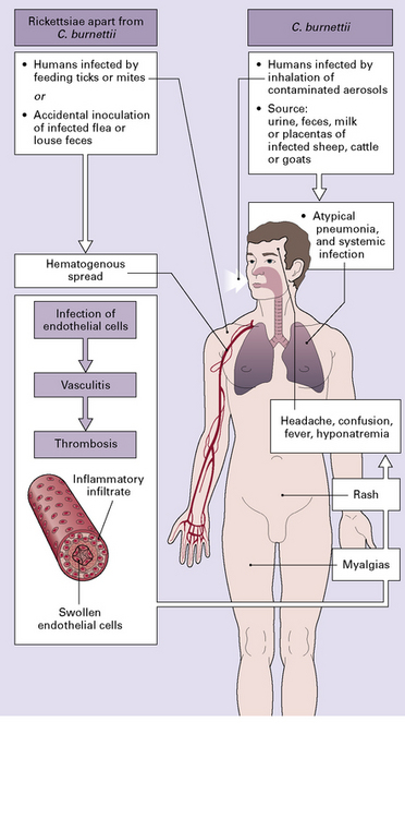
Rickettsiae (apart from C. burnetii) are inoculated into the bloodstream by feeding ticks or mites, or when skin onto which infected flea or louse feces have been deposited is scratched. The rickettsiae are disseminated hematogenously. From the bloodstream they enter, proliferate within, and cause damage to vascular endothelial and smooth muscle cells. Most of the manifestations of human disease (including the spotted fever rash) are a consequence of vasculitis or endothelial involvement with variably severe inflammation (Fig. 14.1). 14.1 Pathogenesis of rickettsial disease. Though vessel wall involvement is considered key to the pathogenesis of rickettsial infections, there is experimental evidence that they can also infect macrophages. Different isolates of even a single microorganism (e.g. R. rickettsii) may induce widely differing degrees of injury in cultured endothelial cells. C. burnetii is present in the urine, feces, milk, and placentas of infected sheep, cattle, and goats. Humans are infected by inhalation of the microorganisms in contaminated aerosols or dust. This results in lung and systemic infection but usually not vasculitis. EPIDEMIOLOGY OF RICKETTSIAL INFECTION Human rickettsial diseases are relatively rare, even in endemic areas. Typhus group infections and Q fever occur worldwide. Most of the other rickettsial diseases are mainly concentrated in specific geographic regions, for example: • Rocky Mountain spotted fever in North, Central, and South America, including the Rocky Mountain region, eastern United States, and Canada • Boutonneuse fever in the Mediterranean region, Asia, and Africa • Queensland tick typhus in northern Australia • scrub typhus in south-east Asia and the Pacific region. A serologic study from Brazil found significant titers of antibodies to Rickettsia rickettsii in as many as 4% of unselected patients in an area endemic for Brazilian spotted fever. HUMAN RICKETTSIAL DISEASES Human rickettsial diseases fall into the following four main groups: • spotted fever: Rocky Mountain spotted fever (R. rickettsii), boutonneuse fever (R. conorii), North Asian tick typhus (R. sibirica), rickettsialpox (R. akari), Queensland tick typhus (R. australis). African tick bite fever (R. africae), North Asian tick typhus (R. sibirica), flea-borne spotted fever (R. felis), oriental spotted fever (R. japonica) • typhus: epidemic typhus (R. prowazekii), endemic/murine typhus (R. typhi) • the microorganism responsible for causing scrub typhus (in south-east Asia and the Pacific region) is now classified in a separate genus of Rickettsiaceae, as Orientia tsutsugamushi • Q fever (Coxiella burnetii).< div class='tao-gold-member'> Only gold members can continue reading. Log In or Register a > to continue Share this:Click to share on Twitter (Opens in new window)Click to share on Facebook (Opens in new window) Related Related posts: Disorders that primarily affect white matter Hyperkinetic movement disorders Neuroepithelial neoplasms displaying neuronal features Multiple sclerosis Acute bacterial infections and bacterial abscesses Fetal and neonatal hypoxic–ischemic lesions Stay updated, free articles. Join our Telegram channel Join Tags: Neuropathology A Reference Text of CNS Pathology Jul 19, 2016 | Posted by admin in NEUROLOGY | Comments Off on Rickettsial and mycoplasma infections Full access? Get Clinical Tree