Fig. 30.1
Schwannoma of the trigeminal nerve. Coronal T1-weighted gadolinium-enhanced MR image. A large enhancing mass is centered in the left parasellar region
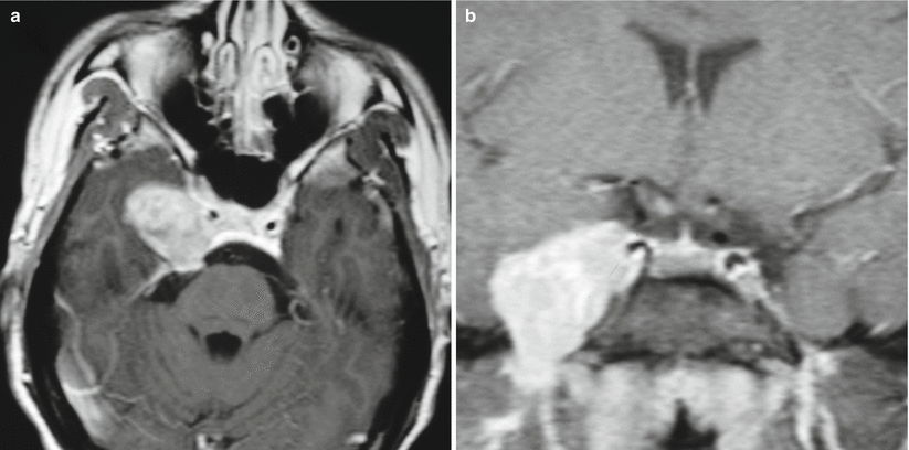
Fig. 30.2
Trigeminal schwannoma. (a) Axial T1-weighted gadolinium-enhanced image. (b) Coronal T1-weighted gadolinium-enhanced image. There is a large right parasellar, enhancing mass centered in the right Meckel’s cave, extending through the right foramen ovale
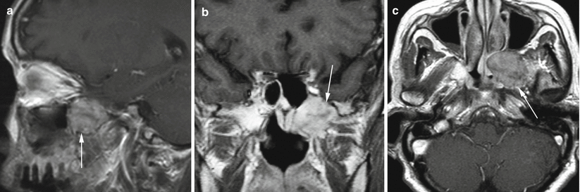
Fig. 30.3
Maxillary nerve schwannoma. (a) Sagittal T1-weighted gadolinium-enhanced image. (b) Coronal T1-weighted gadolinium-enhanced image. (c) Axial T1-weighted gadolinium-enhanced image. There is a heterogeneously enhancing mass (arrows) centered immediately below the left foramen ovale, extending anteriorly to the left pterygopalatine fossa
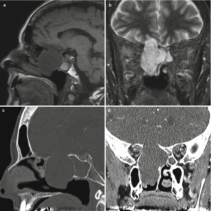
Fig. 30.4
Schwannoma. (a) Sagittal T1-weighted non-enhanced image. (b) Coronal T2-weighted image. (c) Sagittal CT image in bone algorithm. (d) Coronal CT image in soft tissue algorithm. A heterogeneously enhancing mass is centered in the right posterior ethmoid, with marked bony remodeling superiorly, medially, and inferiorly. Superiorly, the mass expands into the right anterior cranial fossa abutting the frontal lobe. Medially, the mass displaces the bony septum and narrows the sphenoid sinuses. Inferiorly, the mass expands into the nasal cavity. The sphenoid sinuses are completely obstructed
30.3 Histopathology
Typical histopathological features of parasellar and sinonasal schwannoma include spindle cell proliferation with low mitotic activity and a low MIB-1 labeling index.
Diffuse immunoreactivity for S100 is typically noted, with negative staining of CD34 and epithelial membrane antigen (EMA) [6].
Classic features of schwannoma—cellular Antoni A and loose Antoni B areas, Verocay bodies, and hyalinized, thickened vessels—are typically observed but not always found in parasellar and sinonasal schwannoma (Fig. 30.5) [7, 8].
Rare, malignant schwannomas with rhabdomyoblastic differentiation (Triton tumors) of the parasellar or sinonasal region have been reported; these tumors require multimodality treatment with radiation and chemotherapy [9].
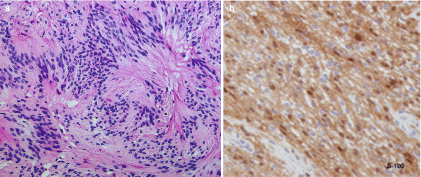
Fig. 30.5
Schwannoma. (a) A schwannoma consisting of dense fascicles in an Antoni A arrangement and nuclear palisading (H&E, ʿÅ ~20), and (b) Differentiated Schwann cells expressing S-100 protein (Adapted with permission from Rallis et al. [8])
30.4 Clinical and Surgical Management
Successful transsphenoidal resection of intrasellar and suprasellar schwannoma has been reported numerous times, typically with improvement in visual function [10, 11].
Endonasal endoscopic approaches may be useful for resection of schwannomas arising in the sinonasal region and more laterally or anteriorly in the skull base, such as the cribriform region, sinonasal tract, or pterygopalatine fossa [12–14].
Schwannomas of the skull base are often characterized by a rich blood supply, making the tumors quite vascular.
For primary cavernous sinus (i.e., trigeminal) schwannomas, open craniotomy with an extradural temporopolar or similar approach may be utilized with excellent surgical results. Facial hypoesthesia is the major adverse outcome associated with this approach [15–17].
30.4.1 Radiotherapy and Radiosurgery
Stereotactic radiosurgery (fractionated or unfractionated) has been reported to achieve successful long-term control of parasellar schwannomas in over 90 % of cases [18–20].Related posts:

Stay updated, free articles. Join our Telegram channel

Full access? Get Clinical Tree


