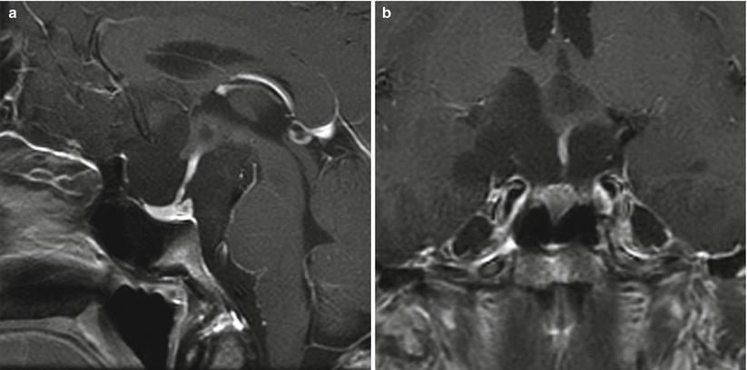
Fig. 26.1
Dermoid cyst. (a) Sagittal T1-weighted post-gadolinium image. (b) Coronal T1-weighted post-gadolinium image. (c) Axial CT image. A lobulated mass is located above the planum sphenoidale anterior to the pituitary stalk. (d) On a CT scan, the lesion has areas of low attenuation indicating the presence of fat. The anterior cerebral arteries are posterior to the lesion

Fig. 26.2
Dermoid cyst. (a) Sagittal T1-weighted noncontrast image. (b) Sagittal T1-weighted post-gadolinium enhanced image. (c) Coronal T1-weighted noncontrast image. Showing a T1 hyperintense heterogeneous and minimally enhancing dermoid cyst of the interpeduncular cisterns posterior to the infundibulum

Fig. 26.3
Epidermoid cyst. (a) Sagittal T1-weighted post-gadolinium image. (b) Coronal T1-weighted post-gadolinium image. There is a cystic lesion in the suprasellar region surrounding the pituitary stalk. The lesion elevates the hypothalami

Fig. 26.4
(a) Sagittal T1-weighted pre-gadolinium image. (b) Coronal T1-weighted pre-gadolinium image. (c) Axial T2-weighted image. A cystic suprasellar lesion contains regions of T1 hyperintensity, possibly representing fat or proteinaceous material. The lesion elevates the left hypothalamus

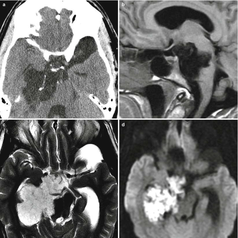
Fig. 26.5
(a) Axial CT scan. (b) Sagittal T1-weighted pre-gadolinium image. (c) Axial T2-weighted image. (d) Axial diffusion-weighted image (DWI). There is a cystic lesion in the suprasellar region, extending to the right ambient cistern and choroidal fissure. There are punctate calcifications along the septated part of the lesion. On DWI, there is increased signal intensity
26.3 Histopathology
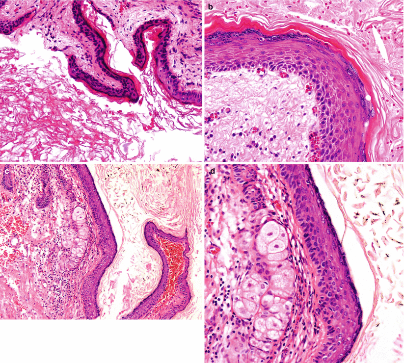
Epidermoid cysts typically exhibit a thin layer of squamous epithelium with a keratohyaline granular layer and a “flaky” keratin formation resting in a fibrocollagenous stroma (Fig. 26.6a, b).
In addition to squamous epithelium, dermoid cysts contain adnexal structures such as hair follicles and glandular elements (Fig. 26.6c, d).
Both cystic sellar lesions (particularly epidermoid cysts) may rupture to cause an exuberant inflammatory reaction in the surrounding parenchyma, resulting in secondary hypophysitis and meningitis.

Fig. 26.6
Epidermoid and dermoid cysts. (a) Low-magnification view of an epidermoid cyst displays the squamous, stratified epithelium lining resting in a fibrous stroma. (b) The epithelium lining shows squamous epithelium with a keratohyaline granular layer and formation of flaky (acellular) keratin. (c, d) Dermoid cysts, on the other hand, show (in addition to the squamous, keratinizing epithelium) adnexal structures beneath the epithelium, including sebaceous glands
26.4 Clinical Management
Surgical resection remains the most effective modality; no adjunctive measures have been proven to be of significant benefit in the management of these lesions.
Extended endonasal microscopic and endoscopic skull base approaches have been utilized for resection of intrasellar and suprasellar dermoid and epidermoid cysts [6, 8, 11, 22–26].
Endoscopy-assisted keyhole approaches may be ideally suited for these lesions because of the ability to look around surgical corners and resect additional cyst contents with the assistance of angled endoscopes [27, 28].
Intraoperatively, dermoid and epidermoid cysts often can prove to be adhesive lesions that are not safely amenable to a gross total resection. The cyst capsule and contents cannot consistently be dissected off key vascular and nervous structures to achieve an acceptable outcome with minimal morbidity. In such cases, conservative resection is recommended.
Steroids are recommended for up to 1 week following rupture or surgery, to reduce the risk of complications from chemical meningitis.
A long-term recurrence rate of 26 % has been reported for all epidermoid and dermoid cysts, mandating serial monitoring of these patients [1].
References
1.
Gormley WB, Tomecek FJ, Qureshi N, Malik GM. Craniocerebral epidermoid and dermoid tumours: a review of 32 cases. Acta Neurochir (Wien). 1994;128:115–21.CrossRef
2.
Sani S, Smith A, Leppla DC, Ilangovan S, Glick R. Epidermoid cyst of the sphenoid sinus with extension into the sella turcica presenting as pituitary apoplexy: case report. Surg Neurol. 2005;63:394–7; discussion 397.CrossRefPubMed
Stay updated, free articles. Join our Telegram channel

Full access? Get Clinical Tree


