Fig. 54.1
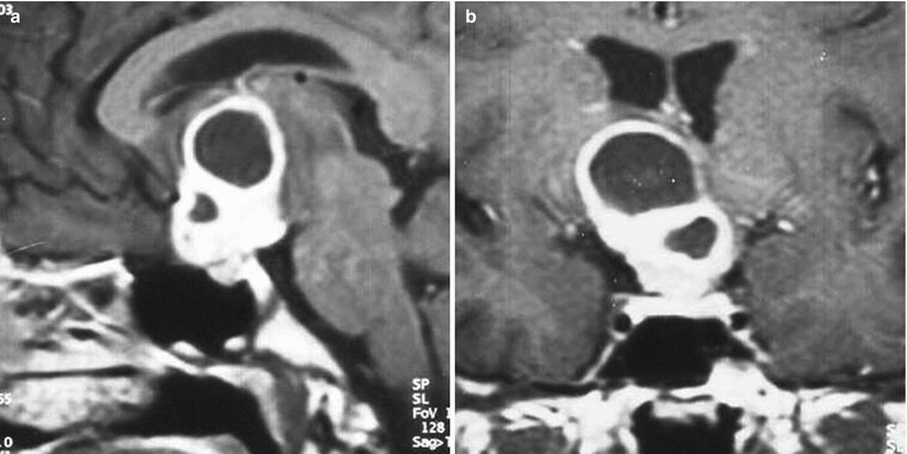
Sellar tuberculoma. (a) Sagittal T1-weighted gadolinium-enhanced image. (b) Coronal T1-weighted gadolinium-enhanced image. A heterogeneously enhancing mass of the sella and suprasellar region is visualized. A patchy area of hypoenhancement is seen in the left aspect of the pituitary gland

Fig. 54.2
Sellar tuberculoma. Sagittal (a) and coronal (b) post-contrast MRI shows a large sellar and suprasellar cystic and solid mass with marked enhancement of the peripheral solid part and a nonenhancing central liquefied area (Adapted from Dutta et al. [7], with permission)
54.3 Histopathology
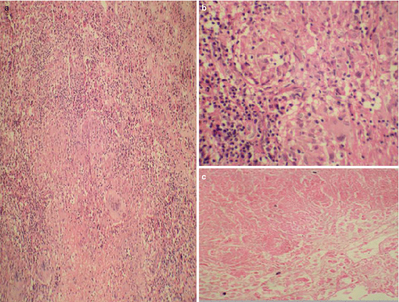
Tuberculous abscesses are characterized by chronic inflammation, granulomatous change, central caseating necrosis, Langhans giant cells, and epithelioid cells (Fig. 54.3).
Polymerase chain reaction (PCR) techniques may aid in the diagnosis of tuberculosis.
CSF analysis may show a lymphocytic reaction with elevated protein.

Fig. 54.3
Tuberculous abscess. Paraffin sections showing multiple epithelioid cell granulomas with Langhans type of giant cell (a) (H&E ×100), epithelioid cells with slipper-shaped nuclei (b) (H&E ×400), and focal areas of necrosis (c) (H&E ×400) (From Furtado et al. [3], with permission)
54.4 Clinical and Surgical Management
Clinical management typically consists of transsphenoidal drainage or resection of the tubercular lesion.
In many cases, dense adhesion to the optic apparatus is noted, and subtotal resection or simple fenestration should be employed.
Following diagnosis and surgical management, patients are placed on long-term antitubercular treatment—typically consisting of rifampin, isoniazid, pyrazinamide, and ethambutol (RIPE therapy)—for 6–18 months.


