Chapter 7 Spinal Column
Spinal Cord and Its Coverings
The surface relationships of the spinal cord and its coverings are of great clinical importance throughout life (Fig. 7.1).

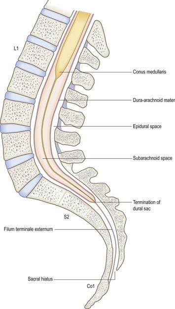
Fig. 7.1 Contents of the vertebral canal in the lumbosacral region.
(Modified with permission from Mackintosh, R.R. 1951. Lumbar Puncture and Spinal Analgesia. E&S Livingstone, Edinburgh.)
During development the vertebral column elongates more rapidly than the spinal cord, which leads to an increasing discrepancy between the anatomical level of spinal cord segments and their corresponding vertebrae. At stage 23, the vertebral column and spinal cord are the same length, and the cord ends at the last coccygeal vertebra; this arrangement continues until the third fetal month. At birth the spinal cord terminates at the lower border of the second lumbar vertebra and may sometimes reach the third lumbar vertebra. In the adult the spinal cord is said to terminate at the level of the disc between the first and second lumbar vertebral bodies, which lies a little above the level of the elbow joint when the arm is by the side and also lies approximately in the transpyloric plane. However, there is considerable variation in the level at which the spinal cord ends. It may end below this level in as many as 40% of subjects, or opposite the body of either the first or second lumbar vertebra; occasionally, it ends opposite the twelfth thoracic or even the third lumbar vertebra.
The dural sac (theca), and thus the subarachnoid space and its contained cerebrospinal fluid (CSF), usually extends to the level of the second segment of the sacrum. This corresponds to the line joining the sacral dimples located in the skin over the posterior superior iliac spines. Occasionally, the dural sac ends as high as the fifth lumbar vertebra, and very rarely it may extend to the third part of the sacrum, in which case it is occasionally possible to enter the subarachnoid space inadvertently during the course of a sacral nerve block.
Clinical Examination
Clinical examination of the back of the trunk and neck best follows the order of inspection, palpation and movement. The examination is determined by the presentation and by the history, and it may include musculoskeletal, neurological and vascular observations. Information relevant to the neurological and vascular examination of the skin and material relating to spinal movements and deeper innervation are presented later. Palpation of the region involves careful assessment of the bony and musculotendinous landmarks described earlier, looking in particular for asymmetry, deformity and tenderness. Note that, apart from the spines, most of the bony elements of the vertebrae and almost all the intervertebral joints are not palpable from behind. In regions of lordosis (sagittal plane curves of the spine with anterior convexity, such as mid-cervical and mid and lower lumbar), parts of the vertebral column can often be palpated anteriorly with care in well-relaxed, thin subjects.
Clinical Procedures
Access to Cerebrospinal Fluid
The safest approach to the CSF is to enter the lumbar cistern of the subarachnoid space in the midline, well below the level at which the spinal cord normally terminates (see Fig. 7.1). The fine needle employed is unlikely to damage the mobile nerve roots of the cauda equina. This procedure is called lumbar puncture. It is also possible to access the CSF by midline puncture of the cerebellomedullary cistern (cisterna magna); this is called cisternal puncture.
Lumbar Puncture: Adult
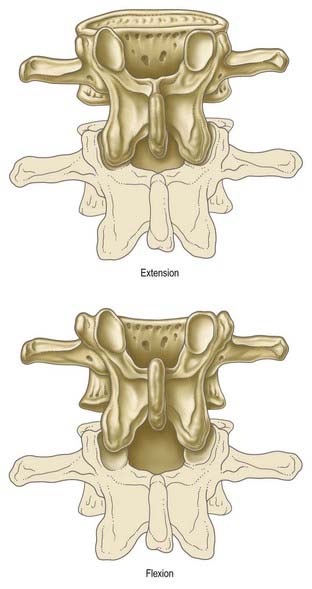
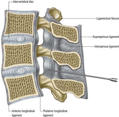
Lumbar puncture in the adult may be performed with the patient either sitting or lying on the side on a firm, flat surface. In each position, the lumbar spine must be flexed as far as possible to separate the vertebral spines maximally and expose the ligamentum flavum in the interlaminar window (Fig. 7.2). A line between the highest points of the iliac crests intersects the vertebral column just above the palpable spine of L4. With the spines now identified, the skin is anaesthetized and a needle is inserted between the spines of L3 and L4 (or L4 and L5). Exact identification of the level by palpation is difficult (Broadbent et al 2000). The soft tissues the needle will ultimately traverse should also be anaesthetized, but care should be taken to avoid injection of an excessive amount of local anaesthetic, which can compromise one’s appreciation of the structures being traversed. These include the subcutaneous fat and supraspinous and interspinous ligaments down to the ligamentum flavum itself. The lumbar puncture needle is then inserted in the midline or just to one side and angled in the horizontal and sagittal planes sufficiently to pierce the ligamentum flavum in or very near the midline (Fig. 7.3).There is a slight loss of resistance as the needle enters the epidural space, and careful advancement pierces the dura and arachnoid to release CSF.
Lumbar Puncture: Neonate and Infant
At full term (40 weeks) the spinal cord usually terminates somewhat lower than the adult level, sometimes reaching the body of L3. The supracristal plane intersects the vertebral column slightly higher (L3–4). By the second postnatal month, the level of cord termination has usually reached its permanent position, level with the body of the first lumbar vertebra. The lower end of the subarachnoid spine is found at sacral level 1 or 2. These differences must be borne in mind when identifying landmarks before undertaking lumbar puncture in neonates and infants.
A lumbar puncture is performed by placing the baby in a position, either lying or ‘sitting,’ that gives maximum convex curvature to the lumbar spine. A needle with trocar is inserted into the back between the spines of the third and fourth lumbar vertebrae and into the subarachnoid space below the level of the conus medullaris. The space between L3 and L4 is approximately level with the iliac crests, and the needle and trocar are usually inserted into the intervertebral space immediately above or below the iliac crests.
Access to the Epidural Space
The epidural ‘space’ lies between the spinal dura and the wall of the vertebral canal. It contains epidural fat and a venous plexus. Access to this space, usually in the lumbar region, is required for the administration of anaesthetic and analgesic drugs and for endoscopy. The caudal route is used mainly for analgesic injections.
Lumbar Epidural
For access to the lumbar epidural space, the approach is as for lumbar puncture. The intention in epidural injection is to avoid dural puncture, so it is best to enter the epidural space in the midline posteriorly, where the depth of the space is greatest. Techniques for entering the epidural space rely on the appreciation of loss of resistance to injection of the chosen medium (usually air or saline) as the space is entered. There is very little distance between the ligamentum flavum and the underlying dura on either side of the median plane.
Caudal Epidural
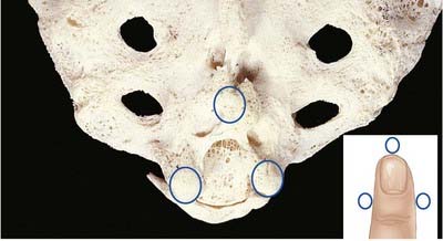
The route of access to the caudal epidural space is via the sacral hiatus. The space is thus entered below the level of termination of the dural sac (S2). With the patient in the lateral position or lying prone over a pelvic pillow, the sacral hiatus is identified by palpation of the sacral cornua (Fig. 7.4). These are felt at the upper end of the natal cleft approximately 5 cm above the tip of the coccyx. Alternatively, the sacral hiatus may be identified by constructing an equilateral triangle based on a line joining the posterior superior iliac spines: the inferior apex of this triangle overlies the hiatus. After local anaesthetic infiltration, a needle is introduced at a 45-degree angle to the skin to penetrate the posterior sacrococcygeal ligament and enter the sacral canal. Once the canal is entered, the hub of the needle is lowered so that the needle may pass along the canal (Fig. 7.5). If the needle is angled too obliquely it will strike bone; if it is placed too superficially it will lie outside the canal. The latter malposition can be confirmed by careful injection of air while palpating the skin over the lower sacrum.

Fig. 7.4 Palpation of the sacral cornua for caudal epidural injection.
(With permission from Ellis, H., Feldman, S.A. 1997. Anatomy for Anaesthetists, 7th ed. Blackwell Science, Oxford.)

Stay updated, free articles. Join our Telegram channel

Full access? Get Clinical Tree

