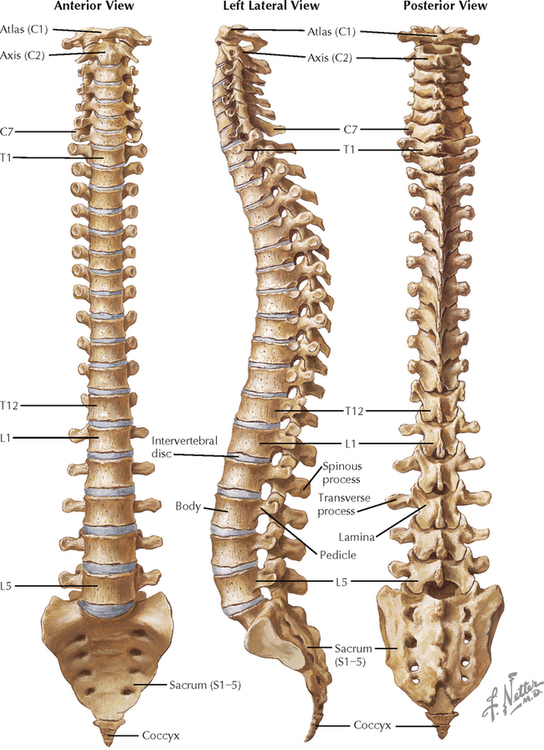
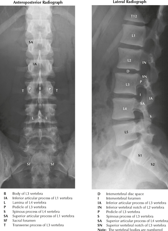
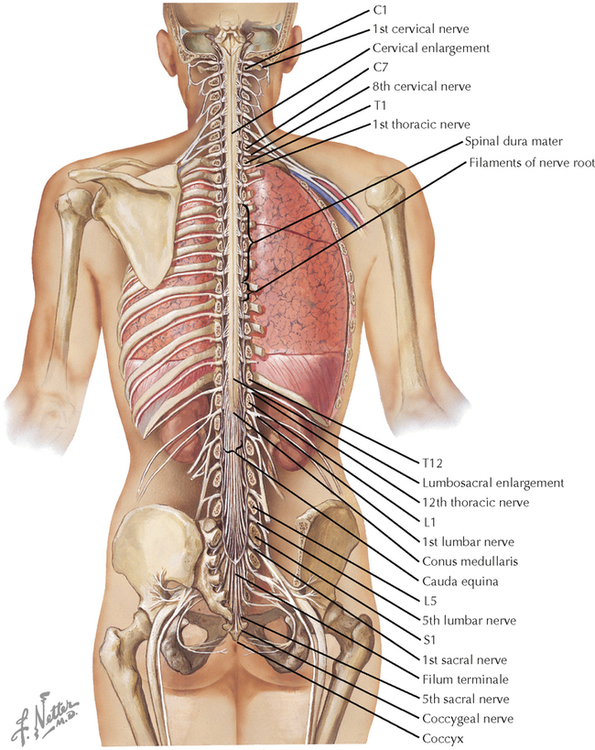
5 SPINAL CORD 5.1. Spinal Column: Bony Anatomy 5.2. Lumbar Vertebrae: Radiography 5.3. Spinal Cord: Gross Anatomy in Situ 5.4. The Spinal Cord: Its Meninges and Spinal Roots 5.5. Spinal Cord: Cross-Sectional Anatomy in Situ 5.6. Spinal Cord: White and Gray Matter 5.1 SPINAL COLUMN: BONY ANATOMY Anterior, lateral, and posterior views of the bony spinal column show the relationships of the intervertebral discs with the vertebral bodies. The discs’ proximity to the intervertebral foramina provides an anatomical substrate for understanding the possible impingement of a herniated nucleus pulposus on spinal roots. Such impingement can cause excruciating, radiating pain if dorsal roots are involved and can cause loss of motor control of affected muscles if ventral roots are involved. In the adult, the spinal cord extends caudally only as far as the L1 vertebral body, leaving the lumbar cistern (the subarachnoid space) accessible for withdrawal of cerebrospinal fluid. 5.2 LUMBAR VERTEBRAE: RADIOGRAPHY These lumbar radiographs show the lumbar spine in an anteroposterior view and a lateral view. The vertebral bodies, with their spinous and transverse processes, are visible, and the spaces occupied by the intervertebral discs are uniform and symmetrical in a normal radiograph. A herniated disc may show a disruption of that symmetry. However, the presence of lumbar radiculopathy and a herniated disc is not always accompanied by radiographic abnormalities. 5.3 SPINAL CORD: GROSS ANATOMY IN SITU The posterior portions of the vertebrae have been removed to show the posterior (dorsal) surface of the spinal cord. Cervical and lumbosacral enlargements of the spinal cord reflect innervation of the limbs. The spinal cord extends rostrally through the foramen magnum, continuous with the medulla. The conus medullaris is located under the L1 vertebral body. The longitudinal growth of the spinal column exceeds that of the spinal cord, causing the spinal cord to end considerably more rostrally in the adult than in the newborn. The associated nerve roots traverse a considerable distance through the subarachnoid space, particularly more caudally in the lumbar cistern, to reach the appropriate intervertebral foramina of exit. In the lumbar cistern, this collection of nerve roots is called the cauda equina (horse’s tail). The lumbar cistern is a large reservoir from which cerebrospinal fluid can be withdrawn. The filum terminale helps to anchor the spinal cord caudally to the coccyx. CLINICAL POINT Only gold members can continue reading. Log In or Register to continue Share this:Click to share on Twitter (Opens in new window)Click to share on Facebook (Opens in new window) Related Related posts: SPINAL CORD MOTOR SYSTEMS NEURONS AND THEIR PROPERTIES AUTONOMIC-HYPOTHALAMIC-LIMBIC SYSTEMS TELENCEPHALON PERIPHERAL NERVOUS SYSTEM Stay updated, free articles. Join our Telegram channel Join Tags: Netters Atlas of Neuroscience with STUDENT CONSULT Online Access Jun 4, 2016 | Posted by admin in NEUROLOGY | Comments Off on SPINAL CORD Full access? Get Clinical Tree
5 SPINAL CORD 5.1. Spinal Column: Bony Anatomy 5.2. Lumbar Vertebrae: Radiography 5.3. Spinal Cord: Gross Anatomy in Situ 5.4. The Spinal Cord: Its Meninges and Spinal Roots 5.5. Spinal Cord: Cross-Sectional Anatomy in Situ 5.6. Spinal Cord: White and Gray Matter 5.1 SPINAL COLUMN: BONY ANATOMY Anterior, lateral, and posterior views of the bony spinal column show the relationships of the intervertebral discs with the vertebral bodies. The discs’ proximity to the intervertebral foramina provides an anatomical substrate for understanding the possible impingement of a herniated nucleus pulposus on spinal roots. Such impingement can cause excruciating, radiating pain if dorsal roots are involved and can cause loss of motor control of affected muscles if ventral roots are involved. In the adult, the spinal cord extends caudally only as far as the L1 vertebral body, leaving the lumbar cistern (the subarachnoid space) accessible for withdrawal of cerebrospinal fluid. 5.2 LUMBAR VERTEBRAE: RADIOGRAPHY These lumbar radiographs show the lumbar spine in an anteroposterior view and a lateral view. The vertebral bodies, with their spinous and transverse processes, are visible, and the spaces occupied by the intervertebral discs are uniform and symmetrical in a normal radiograph. A herniated disc may show a disruption of that symmetry. However, the presence of lumbar radiculopathy and a herniated disc is not always accompanied by radiographic abnormalities. 5.3 SPINAL CORD: GROSS ANATOMY IN SITU The posterior portions of the vertebrae have been removed to show the posterior (dorsal) surface of the spinal cord. Cervical and lumbosacral enlargements of the spinal cord reflect innervation of the limbs. The spinal cord extends rostrally through the foramen magnum, continuous with the medulla. The conus medullaris is located under the L1 vertebral body. The longitudinal growth of the spinal column exceeds that of the spinal cord, causing the spinal cord to end considerably more rostrally in the adult than in the newborn. The associated nerve roots traverse a considerable distance through the subarachnoid space, particularly more caudally in the lumbar cistern, to reach the appropriate intervertebral foramina of exit. In the lumbar cistern, this collection of nerve roots is called the cauda equina (horse’s tail). The lumbar cistern is a large reservoir from which cerebrospinal fluid can be withdrawn. The filum terminale helps to anchor the spinal cord caudally to the coccyx. CLINICAL POINT Only gold members can continue reading. Log In or Register to continue Share this:Click to share on Twitter (Opens in new window)Click to share on Facebook (Opens in new window) Related Related posts: SPINAL CORD MOTOR SYSTEMS NEURONS AND THEIR PROPERTIES AUTONOMIC-HYPOTHALAMIC-LIMBIC SYSTEMS TELENCEPHALON PERIPHERAL NERVOUS SYSTEM Stay updated, free articles. Join our Telegram channel Join Tags: Netters Atlas of Neuroscience with STUDENT CONSULT Online Access Jun 4, 2016 | Posted by admin in NEUROLOGY | Comments Off on SPINAL CORD Full access? Get Clinical Tree