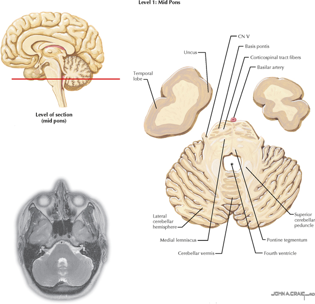
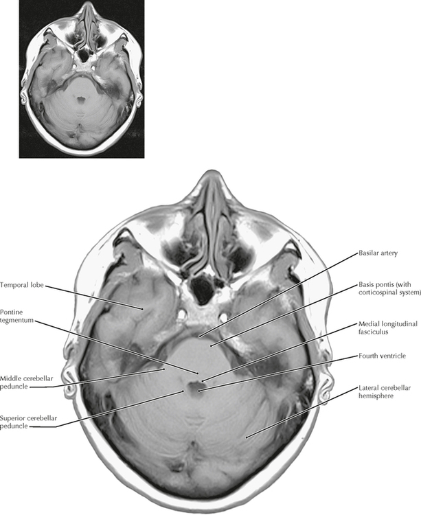
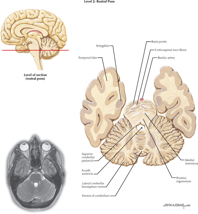
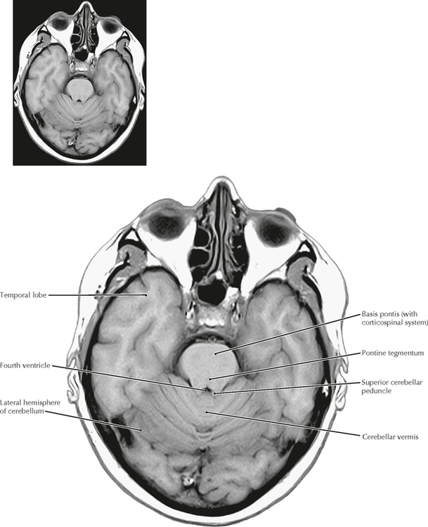
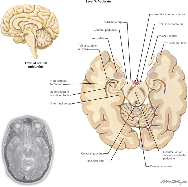
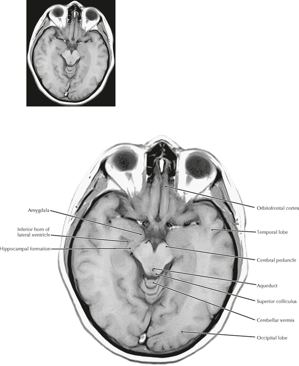
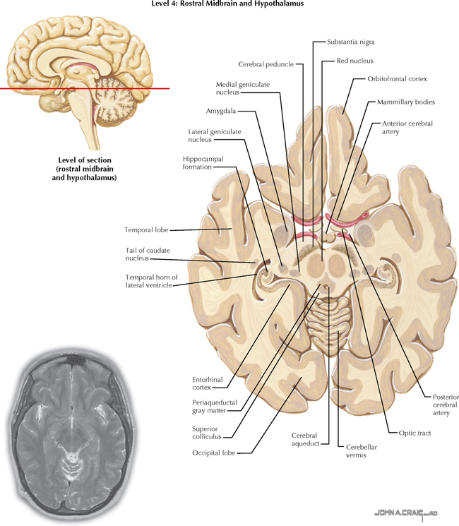
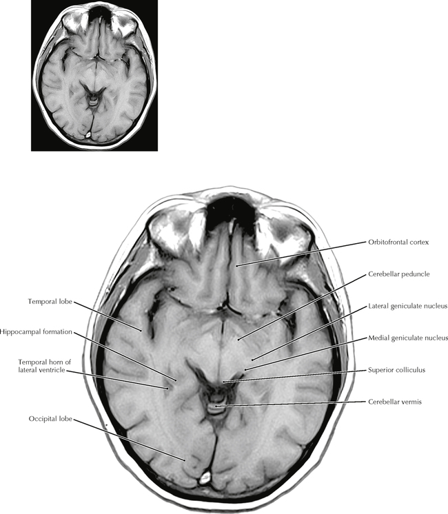
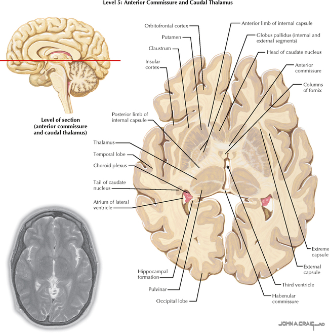
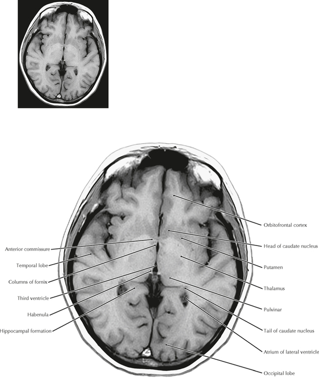
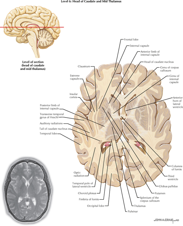
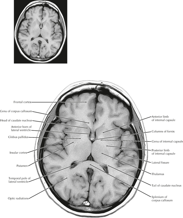
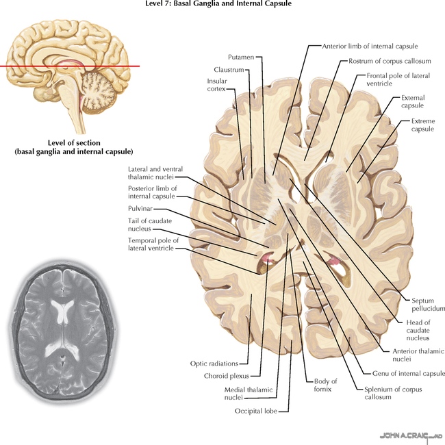
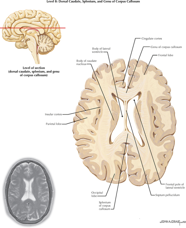
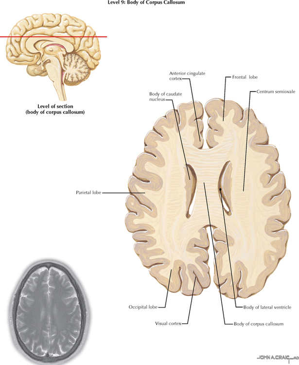
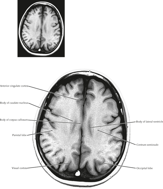
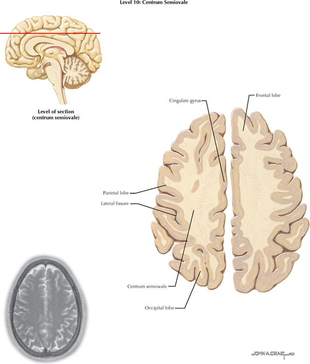
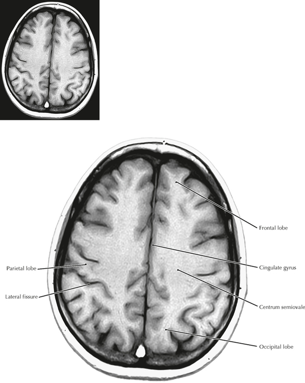
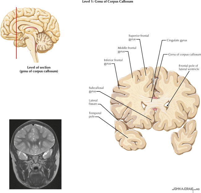
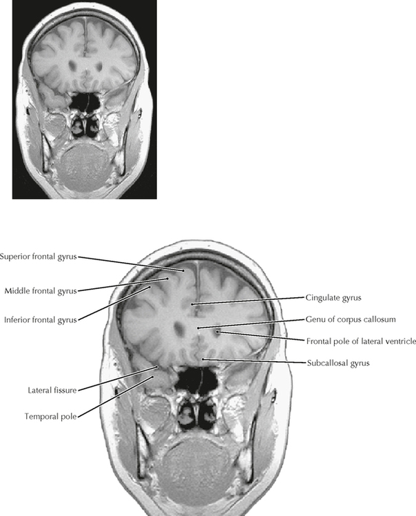
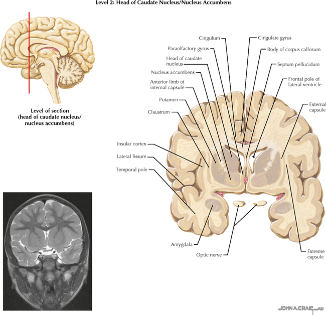
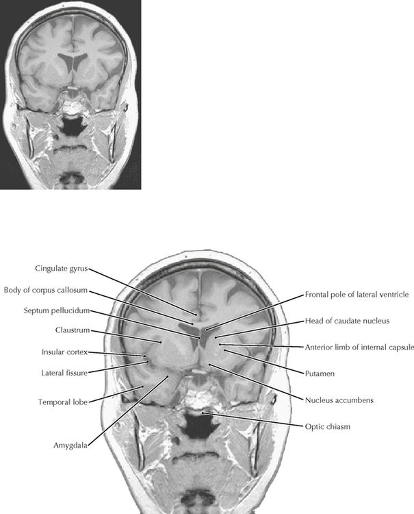
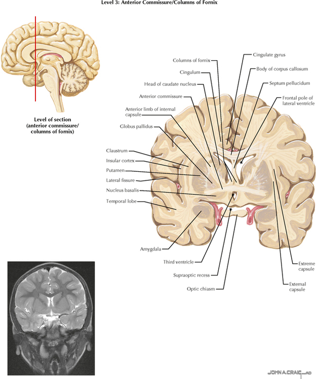
13 TELENCEPHALON 13.1A, B. Axial (Horizontal) Sections Through the Forebrain: Level 1—Mid Pons 13.2A, B. Axial (Horizontal) Sections Through the Forebrain: Level 2—Rostral Pons 13.3A, B. Axial (Horizontal) Sections Through the Forebrain: Level 3—Midbrain 13.4A, B. Axial (Horizontal) Sections Through the Forebrain: Level 4—Rostral Midbrain and Hypothalamus 13.5A, B. Axial (Horizontal) Sections Through the Forebrain: Level 5—Anterior Commissure and Caudal Thalamus 13.6A, B. Axial (Horizontal) Sections Through the Forebrain: Level 6—Head of Caudate and Midthalamus 13.7A, B. Axial (Horizontal) Sections Through the Forebrain: Level 7—Basal Ganglia and Internal Capsule 13.8A, B. Axial (Horizontal) Sections Through the Forebrain: Level 8—Dorsal Caudate, Splenium, and Genu of Corpus Callosum 13.9A, B. Axial (Horizontal) Sections Through the Forebrain: Level 9—Body of Corpus Callosum 13.10A, B. Axial (Horizontal) Sections Through the Forebrain: Level 10—Centrum Semiovale 13.11A, B. Coronal Sections Through the Forebrain: Level 1—Genu of Corpus Callosum 13.12A, B. Coronal Sections Through the Forebrain: Level 2—Head of Caudate Nucleus/Nucleus Accumbens 13.13A, B. Coronal Sections Through the Forebrain: Level 3—Anterior Commissure/Columns of Fornix 13.14A, B. Coronal Sections Through the Forebrain: Level 4—Amygdala, Anterior Limb of Internal Capsule 13.15A, B. Coronal Sections Through the Forebrain: Level 5—Mammillary Body 13.16A, B. Coronal Sections Through the Forebrain: Level 6—Mammillothalamic Tract/Substantia Nigra, Rostral Hippocampus 13.17A, B. Coronal Sections Through the Forebrain: Level 7—Midthalamus 13.18A, B. Coronal Sections Through the Forebrain: Level 8—Geniculate Nuclei 13.19A, B. Coronal Sections Through the Forebrain: Level 9—Caudal Pulvinar and Superior Colliculus 13.20A, B. Coronal Sections Through the Forebrain: Level 10—Splenium of Corpus Callosum 13.21. Layers of the Cerebral Cortex 13.22. Cortical Neuronal Cell Types 13.23. Vertical Columns: Functional Units of the Cerebral Cortex 13.24. Efferent Connections of the Cerebral Cortex 13.25. Neuronal Origins of Efferent Connections of the Cerebral Cortex 13.26. Cortical Association Pathways 13.27. Major Cortical Association Bundles 13.28. Color Imaging of Association Pathways 13.29. Color Imaging of Projection Pathways from the Cerebral Cortex 13.30. Functional Magnetic Resonance Imaging 13.31. Noradrenergic Pathways 13.32. Serotoninergic Pathways 13.33. Dopaminergic Pathways 13.34. Central Cholinergic Pathways 13.35. The Olfactory Nerve and Nerves of the Nose 13.1A AXIAL (HORIZONTAL) SECTIONS THROUGH THE FOREBRAIN: LEVEL 1—MID PONS These axial (horizontal) sections compare anatomical sections and high-resolution magnetic resonance (MR) images. They are cut in the true horizontal (axial) plane, not in the older 25-degree tilt. The most important anatomical relationships in these sections center on the internal capsule (IC). The head of the caudate nucleus is medial to the anterior limb of the IC and forms the lateral margin of the frontal pole of the lateral ventricle. The thalamus is medial to the posterior limb of the IC. The globus pallidus and putamen are lateral to the wedge-shaped IC. The posterior limb of the IC carries the major descending corticospinal, corticorubral, and corticoreticular fibers and the ascending sensory fibers of the somatosensory and trigeminal systems. The most posterior portions of the posterior limb also carry the auditory and visual projections to their respective cortices. The genu of the IC carries the corticobulbar fibers. The anterior limb of the IC carries cortical projections to the striatum and the pontine nuclei (pontocerebellar system). The full-plate MR images are T1-weighted; the ventricles appear dark. The scout MRI images that accompany the drawings are T2-weighted MR images, in which the CSF appears white. 13.1B AXIAL (HORIZONTAL) SECTIONS THROUGH THE FOREBRAIN: LEVEL 1—MID PONS 13.2A AXIAL (HORIZONTAL) SECTIONS THROUGH THE FOREBRAIN: LEVEL 2—ROSTRAL PONS 13.2B AXIAL (HORIZONTAL) SECTIONS THROUGH THE FOREBRAIN: LEVEL 2—ROSTRAL PONS 13.3A AXIAL (HORIZONTAL) SECTIONS THROUGH THE FOREBRAIN: LEVEL 3—MIDBRAIN CLINICAL POINT The temporal lobe includes the amygdaloid nuclei, the hippocampal formation and associated cortex, the transverse gyrus of Heschl, some language-associated cortical regions (Wernicke’s area in the dominant hemisphere), Meyer’s loop of geniculocalcarine axons, the inferior horn of the lateral ventricle, and extensive cortical areas (superior, middle, and inferior temporal gyri). The temporal lobe can be damaged by trauma, infarcts, tumors, abscesses, and other pathological conditions. Such damage can result in auditory hallucinations, delirium and psychotic behavior, sometimes a contralateral upper quadrantanopia (if Meyer’s loop is damaged), and receptive aphasia (Wernicke’s aphasia) that involves a lack of understanding of verbal information (in a lesion of the dominant hemisphere). Some very specific lesions in the temporal lobe result in an agnosia for recognition of faces (prosopagnosia). 13.3B AXIAL (HORIZONTAL) SECTIONS THROUGH THE FOREBRAIN: LEVEL 3—MIDBRAIN 13.4A AXIAL (HORIZONTAL) SECTIONS THROUGH THE FOREBRAIN: LEVEL 4—ROSTRAL MIDBRAIN AND HYPOTHALAMUS 13.4B AXIAL (HORIZONTAL) SECTIONS THROUGH THE FOREBRAIN: LEVEL 4—ROSTRAL MIDBRAIN AND HYPOTHALAMUS 13.5A AXIAL (HORIZONTAL) SECTIONS THROUGH THE FOREBRAIN: LEVEL 5—ANTERIOR COMMISSURE AND CAUDAL THALAMUS CLINICAL POINT The basal ganglia assist the cerebral cortex in planning and generating desired programs of activity and suppressing undesired programs of activity. The most conspicuous arena in which these functions are observed is motor activity. Basal ganglia disorders produce movement problems that are often involuntary in nature and are commonly accompanied by cognitive and affective symptoms (e.g., Huntington’s disease). The principal route of information flow from the basal ganglia is from the thalamus and cerebral cortex to the striatum (caudate nucleus and putamen), then to the globus pallidus, then back to the thalamus and cortex, completing the loop. Disruption of this loop can produce excessive movements (e.g., choreiform and athetoid movements, tremor) or diminished movements (bradykinesia). In some instances, specific nuclei are known to be associated with such changes. A small lacunar infarct in the subthalamic nucleus results in wild, flinging (ballistic) movements in the contralateral limbs. However, a surgical lesion in the subthalamic nucleus may ameliorate some of the movement problems seen in Parkinson’s disease. The subthalamus most likely drives activity in the internal segment of the globus pallidus, which in turn can be modified by the external segment. A pathological lesion in the globus pallidus can produce rigidity and akinesia; a surgical pallidal lesion may reduce excessive movements in other basal ganglia disorders. 13.5B AXIAL (HORIZONTAL) SECTIONS THROUGH THE FOREBRAIN: LEVEL 5—ANTERIOR COMMISSURE AND CAUDAL THALAMUS 13.6A AXIAL (HORIZONTAL) SECTIONS THROUGH THE FOREBRAIN: LEVEL 6—HEAD OF CAUDATE AND MID THALAMUS 13.6B AXIAL (HORIZONTAL) SECTIONS THROUGH THE FOREBRAIN: LEVEL 6—HEAD OF CAUDATE AND MID THALAMUS 13.7A AXIAL (HORIZONTAL) SECTIONS THROUGH THE FOREBRAIN: LEVEL 7—BASAL GANGLIA AND INTERNAL CAPSULE CLINICAL POINT Huntington’s disease is an autosomal dominant disorder caused by a trinucleotide repeat (CAG) on the short arm of chromosome 4. It results in a progressive, untreatable disease that includes a movement disorder (choreiform movements: brisk, jerky, forcible, arrhythmic movements), progressive cognitive impairment, and affective disorders (such as depression, psychotic behavior). This disease progresses from a state of minor impairment (clumsiness) with minor behavioral problems (irritability and depression) to major impairment, dementia, and a decline that leads to incapacitation and ultimately to an early death. The anatomical hallmark of this disease is marked degeneration of the caudate nucleus (also the putamen). The characteristic bulge of the head of the caudate into the frontal pole of the lateral ventricle is lost. Most of the medium spiny caudate neurons that project to the globus pallidus degenerate as the result of damage from excess Ca++ influx caused by glutamate excitotoxic damage via activation of receptors of N-methyl-d-aspartate (NMDA). The intrinsic cholinergic interneurons of the striatum also degenerate in Huntington’s disease. 13.7B AXIAL (HORIZONTAL) SECTIONS THROUGH THE FOREBRAIN: LEVEL 7—BASAL GANGLIA AND INTERNAL CAPSULE 13.8A AXIAL (HORIZONTAL) SECTIONS THROUGH THE FOREBRAIN: LEVEL 8—DORSAL CAUDATE, SPLENIUM, AND GENU OF CORPUS CALLOSUM 13.8B AXIAL (HORIZONTAL) SECTIONS THROUGH THE FOREBRAIN: LEVEL 8—DORSAL CAUDATE, SPLENIUM, AND GENU OF CORPUS CALLOSUM 13.9A AXIAL (HORIZONTAL) SECTIONS THROUGH THE FOREBRAIN: LEVEL 9—BODY OF CORPUS CALLOSUM 13.9B AXIAL (HORIZONTAL) SECTIONS THROUGH THE FOREBRAIN: LEVEL 9—BODY OF CORPUS CALLOSUM 13.10A AXIAL (HORIZONTAL) SECTIONS THROUGH THE FOREBRAIN: LEVEL 10—CENTRUM SEMIOVALE 13.10B AXIAL (HORIZONTAL) SECTIONS THROUGH THE FOREBRAIN: LEVEL 10—CENTRUM SEMIOVALE 13.11A CORONAL SECTIONS THROUGH THE FOREBRAIN: LEVEL 1—GENU OF CORPUS CALLOSUM These coronal sections compare anatomical sections and high-resolution MR images. They show important relationships among the internal capsule, basal ganglia, and thalamus. These sections show basal forebrain structures, such as nucleus accumbens, substantia innominata, and nucleus basalis (cholinergic forebrain nucleus), some individual thalamic nuclei, and the important temporal lobe structures (amygdaloid nuclei, hippocampal formation) and pathways (fornix, stria terminalis). The full-page MR images are T1-weighted; the ventricles appear dark. The scout MR images that accompany the drawings are T2-weighted MR images in which the CSF appears white. 13.11B CORONAL SECTIONS THROUGH THE FOREBRAIN: LEVEL 1—GENU OF CORPUS CALLOSUM 13.12A CORONAL SECTIONS THROUGH THE FOREBRAIN: LEVEL 2—HEAD OF CAUDATE NUCLEUS/NUCLEUS ACCUMBENS CLINICAL POINT The nucleus accumbens is located at the anterior end of the striatum in the ventral part of the forebrain. It receives a variety of inputs from limbic structures, such as the amygdala, hypocampal formation, and bed nucleus of the stria terminalis. A major dopaminergic (DA) input innervates the nucleus accumbens via the mesolimbic DA pathway, which derives from the ventral tegmental area in the ventral midbrain. The nucleus accumbens is central to motivational states and addictive behavior, driven by DA neurotransmission. The nucleus accumbens is also a principal region of brain circuitry associated with reward, such as joy, pleasure, and gratification. This nucleus has a looped circuitry through the thalamus and cortex that helps to provide motor expression of emotional responses and accompanying gestures and behaviors. 13.12B CORONAL SECTIONS THROUGH THE FOREBRAIN: LEVEL 2—HEAD OF CAUDATE NUCLEUS/NUCLEUS ACCUMBENS 13.13A Only gold members can continue reading. Log In or Register to continue Share this:Click to share on Twitter (Opens in new window)Click to share on Facebook (Opens in new window) Related Related posts: VENTRICLES AND THE CEREBROSPINAL FLUID SPINAL CORD MOTOR SYSTEMS SENSORY SYSTEMS AUTONOMIC-HYPOTHALAMIC-LIMBIC SYSTEMS PERIPHERAL NERVOUS SYSTEM Stay updated, free articles. Join our Telegram channel Join Tags: Netters Atlas of Neuroscience with STUDENT CONSULT Online Access Jun 4, 2016 | Posted by admin in NEUROLOGY | Comments Off on TELENCEPHALON Full access? Get Clinical Tree
13 TELENCEPHALON 13.1A, B. Axial (Horizontal) Sections Through the Forebrain: Level 1—Mid Pons 13.2A, B. Axial (Horizontal) Sections Through the Forebrain: Level 2—Rostral Pons 13.3A, B. Axial (Horizontal) Sections Through the Forebrain: Level 3—Midbrain 13.4A, B. Axial (Horizontal) Sections Through the Forebrain: Level 4—Rostral Midbrain and Hypothalamus 13.5A, B. Axial (Horizontal) Sections Through the Forebrain: Level 5—Anterior Commissure and Caudal Thalamus 13.6A, B. Axial (Horizontal) Sections Through the Forebrain: Level 6—Head of Caudate and Midthalamus 13.7A, B. Axial (Horizontal) Sections Through the Forebrain: Level 7—Basal Ganglia and Internal Capsule 13.8A, B. Axial (Horizontal) Sections Through the Forebrain: Level 8—Dorsal Caudate, Splenium, and Genu of Corpus Callosum 13.9A, B. Axial (Horizontal) Sections Through the Forebrain: Level 9—Body of Corpus Callosum 13.10A, B. Axial (Horizontal) Sections Through the Forebrain: Level 10—Centrum Semiovale 13.11A, B. Coronal Sections Through the Forebrain: Level 1—Genu of Corpus Callosum 13.12A, B. Coronal Sections Through the Forebrain: Level 2—Head of Caudate Nucleus/Nucleus Accumbens 13.13A, B. Coronal Sections Through the Forebrain: Level 3—Anterior Commissure/Columns of Fornix 13.14A, B. Coronal Sections Through the Forebrain: Level 4—Amygdala, Anterior Limb of Internal Capsule 13.15A, B. Coronal Sections Through the Forebrain: Level 5—Mammillary Body 13.16A, B. Coronal Sections Through the Forebrain: Level 6—Mammillothalamic Tract/Substantia Nigra, Rostral Hippocampus 13.17A, B. Coronal Sections Through the Forebrain: Level 7—Midthalamus 13.18A, B. Coronal Sections Through the Forebrain: Level 8—Geniculate Nuclei 13.19A, B. Coronal Sections Through the Forebrain: Level 9—Caudal Pulvinar and Superior Colliculus 13.20A, B. Coronal Sections Through the Forebrain: Level 10—Splenium of Corpus Callosum 13.21. Layers of the Cerebral Cortex 13.22. Cortical Neuronal Cell Types 13.23. Vertical Columns: Functional Units of the Cerebral Cortex 13.24. Efferent Connections of the Cerebral Cortex 13.25. Neuronal Origins of Efferent Connections of the Cerebral Cortex 13.26. Cortical Association Pathways 13.27. Major Cortical Association Bundles 13.28. Color Imaging of Association Pathways 13.29. Color Imaging of Projection Pathways from the Cerebral Cortex 13.30. Functional Magnetic Resonance Imaging 13.31. Noradrenergic Pathways 13.32. Serotoninergic Pathways 13.33. Dopaminergic Pathways 13.34. Central Cholinergic Pathways 13.35. The Olfactory Nerve and Nerves of the Nose 13.1A AXIAL (HORIZONTAL) SECTIONS THROUGH THE FOREBRAIN: LEVEL 1—MID PONS These axial (horizontal) sections compare anatomical sections and high-resolution magnetic resonance (MR) images. They are cut in the true horizontal (axial) plane, not in the older 25-degree tilt. The most important anatomical relationships in these sections center on the internal capsule (IC). The head of the caudate nucleus is medial to the anterior limb of the IC and forms the lateral margin of the frontal pole of the lateral ventricle. The thalamus is medial to the posterior limb of the IC. The globus pallidus and putamen are lateral to the wedge-shaped IC. The posterior limb of the IC carries the major descending corticospinal, corticorubral, and corticoreticular fibers and the ascending sensory fibers of the somatosensory and trigeminal systems. The most posterior portions of the posterior limb also carry the auditory and visual projections to their respective cortices. The genu of the IC carries the corticobulbar fibers. The anterior limb of the IC carries cortical projections to the striatum and the pontine nuclei (pontocerebellar system). The full-plate MR images are T1-weighted; the ventricles appear dark. The scout MRI images that accompany the drawings are T2-weighted MR images, in which the CSF appears white. 13.1B AXIAL (HORIZONTAL) SECTIONS THROUGH THE FOREBRAIN: LEVEL 1—MID PONS 13.2A AXIAL (HORIZONTAL) SECTIONS THROUGH THE FOREBRAIN: LEVEL 2—ROSTRAL PONS 13.2B AXIAL (HORIZONTAL) SECTIONS THROUGH THE FOREBRAIN: LEVEL 2—ROSTRAL PONS 13.3A AXIAL (HORIZONTAL) SECTIONS THROUGH THE FOREBRAIN: LEVEL 3—MIDBRAIN CLINICAL POINT The temporal lobe includes the amygdaloid nuclei, the hippocampal formation and associated cortex, the transverse gyrus of Heschl, some language-associated cortical regions (Wernicke’s area in the dominant hemisphere), Meyer’s loop of geniculocalcarine axons, the inferior horn of the lateral ventricle, and extensive cortical areas (superior, middle, and inferior temporal gyri). The temporal lobe can be damaged by trauma, infarcts, tumors, abscesses, and other pathological conditions. Such damage can result in auditory hallucinations, delirium and psychotic behavior, sometimes a contralateral upper quadrantanopia (if Meyer’s loop is damaged), and receptive aphasia (Wernicke’s aphasia) that involves a lack of understanding of verbal information (in a lesion of the dominant hemisphere). Some very specific lesions in the temporal lobe result in an agnosia for recognition of faces (prosopagnosia). 13.3B AXIAL (HORIZONTAL) SECTIONS THROUGH THE FOREBRAIN: LEVEL 3—MIDBRAIN 13.4A AXIAL (HORIZONTAL) SECTIONS THROUGH THE FOREBRAIN: LEVEL 4—ROSTRAL MIDBRAIN AND HYPOTHALAMUS 13.4B AXIAL (HORIZONTAL) SECTIONS THROUGH THE FOREBRAIN: LEVEL 4—ROSTRAL MIDBRAIN AND HYPOTHALAMUS 13.5A AXIAL (HORIZONTAL) SECTIONS THROUGH THE FOREBRAIN: LEVEL 5—ANTERIOR COMMISSURE AND CAUDAL THALAMUS CLINICAL POINT The basal ganglia assist the cerebral cortex in planning and generating desired programs of activity and suppressing undesired programs of activity. The most conspicuous arena in which these functions are observed is motor activity. Basal ganglia disorders produce movement problems that are often involuntary in nature and are commonly accompanied by cognitive and affective symptoms (e.g., Huntington’s disease). The principal route of information flow from the basal ganglia is from the thalamus and cerebral cortex to the striatum (caudate nucleus and putamen), then to the globus pallidus, then back to the thalamus and cortex, completing the loop. Disruption of this loop can produce excessive movements (e.g., choreiform and athetoid movements, tremor) or diminished movements (bradykinesia). In some instances, specific nuclei are known to be associated with such changes. A small lacunar infarct in the subthalamic nucleus results in wild, flinging (ballistic) movements in the contralateral limbs. However, a surgical lesion in the subthalamic nucleus may ameliorate some of the movement problems seen in Parkinson’s disease. The subthalamus most likely drives activity in the internal segment of the globus pallidus, which in turn can be modified by the external segment. A pathological lesion in the globus pallidus can produce rigidity and akinesia; a surgical pallidal lesion may reduce excessive movements in other basal ganglia disorders. 13.5B AXIAL (HORIZONTAL) SECTIONS THROUGH THE FOREBRAIN: LEVEL 5—ANTERIOR COMMISSURE AND CAUDAL THALAMUS 13.6A AXIAL (HORIZONTAL) SECTIONS THROUGH THE FOREBRAIN: LEVEL 6—HEAD OF CAUDATE AND MID THALAMUS 13.6B AXIAL (HORIZONTAL) SECTIONS THROUGH THE FOREBRAIN: LEVEL 6—HEAD OF CAUDATE AND MID THALAMUS 13.7A AXIAL (HORIZONTAL) SECTIONS THROUGH THE FOREBRAIN: LEVEL 7—BASAL GANGLIA AND INTERNAL CAPSULE CLINICAL POINT Huntington’s disease is an autosomal dominant disorder caused by a trinucleotide repeat (CAG) on the short arm of chromosome 4. It results in a progressive, untreatable disease that includes a movement disorder (choreiform movements: brisk, jerky, forcible, arrhythmic movements), progressive cognitive impairment, and affective disorders (such as depression, psychotic behavior). This disease progresses from a state of minor impairment (clumsiness) with minor behavioral problems (irritability and depression) to major impairment, dementia, and a decline that leads to incapacitation and ultimately to an early death. The anatomical hallmark of this disease is marked degeneration of the caudate nucleus (also the putamen). The characteristic bulge of the head of the caudate into the frontal pole of the lateral ventricle is lost. Most of the medium spiny caudate neurons that project to the globus pallidus degenerate as the result of damage from excess Ca++ influx caused by glutamate excitotoxic damage via activation of receptors of N-methyl-d-aspartate (NMDA). The intrinsic cholinergic interneurons of the striatum also degenerate in Huntington’s disease. 13.7B AXIAL (HORIZONTAL) SECTIONS THROUGH THE FOREBRAIN: LEVEL 7—BASAL GANGLIA AND INTERNAL CAPSULE 13.8A AXIAL (HORIZONTAL) SECTIONS THROUGH THE FOREBRAIN: LEVEL 8—DORSAL CAUDATE, SPLENIUM, AND GENU OF CORPUS CALLOSUM 13.8B AXIAL (HORIZONTAL) SECTIONS THROUGH THE FOREBRAIN: LEVEL 8—DORSAL CAUDATE, SPLENIUM, AND GENU OF CORPUS CALLOSUM 13.9A AXIAL (HORIZONTAL) SECTIONS THROUGH THE FOREBRAIN: LEVEL 9—BODY OF CORPUS CALLOSUM 13.9B AXIAL (HORIZONTAL) SECTIONS THROUGH THE FOREBRAIN: LEVEL 9—BODY OF CORPUS CALLOSUM 13.10A AXIAL (HORIZONTAL) SECTIONS THROUGH THE FOREBRAIN: LEVEL 10—CENTRUM SEMIOVALE 13.10B AXIAL (HORIZONTAL) SECTIONS THROUGH THE FOREBRAIN: LEVEL 10—CENTRUM SEMIOVALE 13.11A CORONAL SECTIONS THROUGH THE FOREBRAIN: LEVEL 1—GENU OF CORPUS CALLOSUM These coronal sections compare anatomical sections and high-resolution MR images. They show important relationships among the internal capsule, basal ganglia, and thalamus. These sections show basal forebrain structures, such as nucleus accumbens, substantia innominata, and nucleus basalis (cholinergic forebrain nucleus), some individual thalamic nuclei, and the important temporal lobe structures (amygdaloid nuclei, hippocampal formation) and pathways (fornix, stria terminalis). The full-page MR images are T1-weighted; the ventricles appear dark. The scout MR images that accompany the drawings are T2-weighted MR images in which the CSF appears white. 13.11B CORONAL SECTIONS THROUGH THE FOREBRAIN: LEVEL 1—GENU OF CORPUS CALLOSUM 13.12A CORONAL SECTIONS THROUGH THE FOREBRAIN: LEVEL 2—HEAD OF CAUDATE NUCLEUS/NUCLEUS ACCUMBENS CLINICAL POINT The nucleus accumbens is located at the anterior end of the striatum in the ventral part of the forebrain. It receives a variety of inputs from limbic structures, such as the amygdala, hypocampal formation, and bed nucleus of the stria terminalis. A major dopaminergic (DA) input innervates the nucleus accumbens via the mesolimbic DA pathway, which derives from the ventral tegmental area in the ventral midbrain. The nucleus accumbens is central to motivational states and addictive behavior, driven by DA neurotransmission. The nucleus accumbens is also a principal region of brain circuitry associated with reward, such as joy, pleasure, and gratification. This nucleus has a looped circuitry through the thalamus and cortex that helps to provide motor expression of emotional responses and accompanying gestures and behaviors. 13.12B CORONAL SECTIONS THROUGH THE FOREBRAIN: LEVEL 2—HEAD OF CAUDATE NUCLEUS/NUCLEUS ACCUMBENS 13.13A Only gold members can continue reading. Log In or Register to continue Share this:Click to share on Twitter (Opens in new window)Click to share on Facebook (Opens in new window) Related Related posts: VENTRICLES AND THE CEREBROSPINAL FLUID SPINAL CORD MOTOR SYSTEMS SENSORY SYSTEMS AUTONOMIC-HYPOTHALAMIC-LIMBIC SYSTEMS PERIPHERAL NERVOUS SYSTEM Stay updated, free articles. Join our Telegram channel Join Tags: Netters Atlas of Neuroscience with STUDENT CONSULT Online Access Jun 4, 2016 | Posted by admin in NEUROLOGY | Comments Off on TELENCEPHALON Full access? Get Clinical Tree