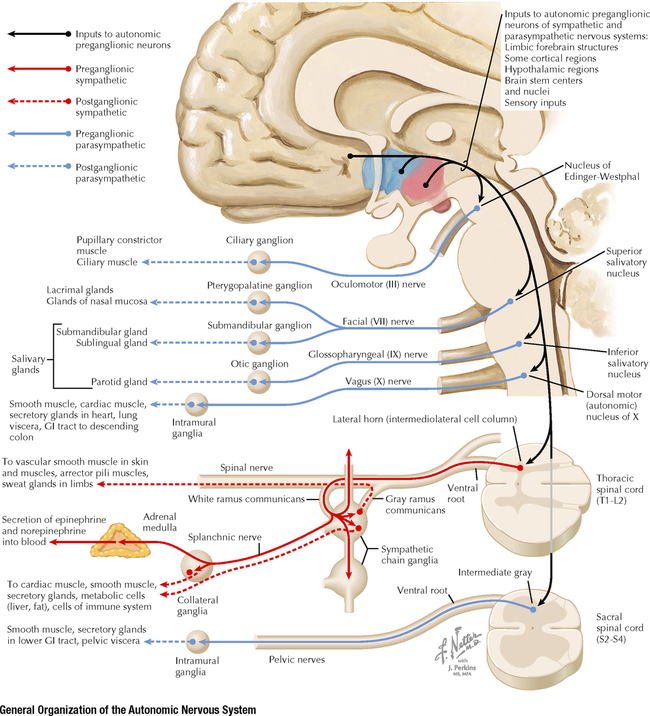
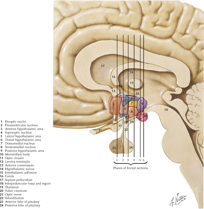
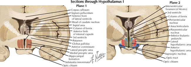
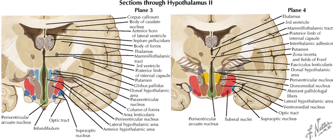
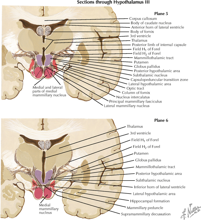
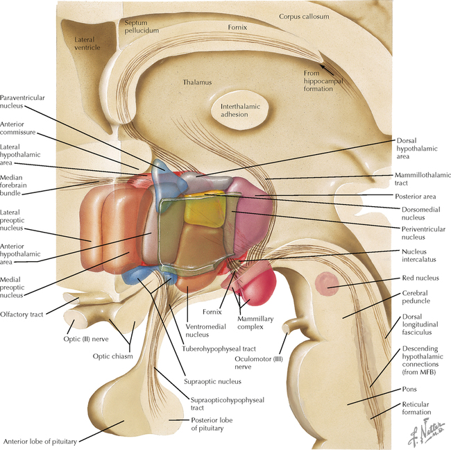
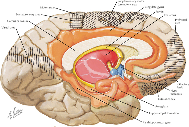
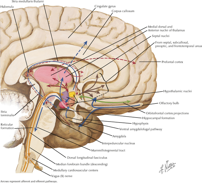
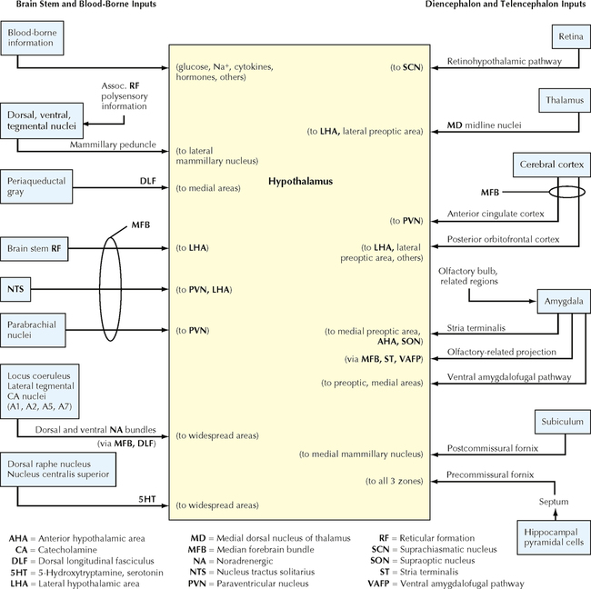
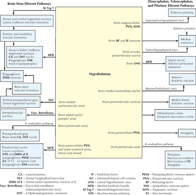
16 AUTONOMIC-HYPOTHALAMIC-LIMBIC SYSTEMS Autonomic Nervous System 16.1. General Organization of the Autonomic Nervous System Hypothalamus and Pituitary 16.2. General Anatomy of the Hypothalamus 16.3. Sections through the Hypothalamus: Preoptic and Supraoptic Zones 16.4. Sections through the Hypothalamus: Tuberal Zone 16.5. Sections through the Hypothalamus: Mammillary Zone 16.6. Schematic Reconstruction of the Hypothalamus 16.7. Forebrain Regions Associated with the Hypothalamus 16.8. Afferent and Efferent Pathways Associated with the Hypothalamus 16.9. Schematic Diagram of Major Hypothalamic Afferent Pathways 16.10. Schematic Diagram of Major Hypothalamic Efferent Pathways 16.11. Summary of General Hypothalamic Connections 16.12. Paraventricular Nucleus of the Hypothalamus: Regulation of Pituitary Neurohormonal Outflow, Autonomic Preganglionic Outflow, and Limbic Activity 16.13. Mechanisms of Cytokine Influences on the Hypothalamus and Other Brain Regions and on Behavior 16.14. Circumventricular Organs 16.15. The Hypophyseal Portal Vasculature 16.16. Regulation of Anterior Pituitary Hormone Secretion 16.17. Posterior Pituitary (Neurohypophyseal) Hormones: Oxytocin and Vasopressin 16.18. Vasopressin (Antidiuretic Hormone) Regulation of Water Balance and Fluid Osmolality 16.19. The Hypothalamus and Thermoregulation 16.20. Short-Term Regulation of Blood Pressure 16.21. Long-Term Regulation of Blood Pressure 16.22. Neural Control of Appetite and Hunger 16.23. Neural and Neuroendocrine Roles in the Fight-or-Flight Response 16.24. Neuroimmunomodulation Limbic System 16.25. Anatomy of the Limbic Forebrain 16.26. Hippocampal Formation: General Anatomy 16.27. Neuronal Connections in the Hippocampal Formation 16.28. Major Afferent and Efferent Connections of the Hippocampal Formation 16.29. Major Afferent Connections of the Amygdala 16.30. Major Efferent Connections of the Amygdala 16.31. Summary of Major Afferents, Efferents, and Interconnections of the Amygdala 16.32. Major Afferent and Efferent Connections of the Septal Nuclei 16.33. Major Connections of the Cingulate Cortex Olfactory System 16.34. Olfactory Receptors 16.35. Olfactory Pathways AUTONOMIC NERVOUS SYSTEM 16.1 GENERAL ORGANIZATION OF THE AUTONOMIC NERVOUS SYSTEM The autonomic nervous system is a two-neuron chain connecting preganglionic neurons through ganglia to visceral target tissues (cardiac muscle, smooth muscle, secretory glands, metabolic cells, cells of the immune system). The sympathetic division (sympathetic nervous system; SNS) is a thoracolumbar (T1–L2) system arising from the intermediolateral cell column of the lateral horn of the spinal cord, acting through chain ganglia and collateral ganglia; it is a system designed for fight-or-flight reactions in an emergency. The parasympathetic division (parasympathetic nervous system) is a craniosacral system arising from brain stem nuclei associated with cranial nerves (CNs) III, VII, IX, and X and from the intermediate gray in the S2–S4 spinal cord. Connections from CNs III, VII, and IX act through cranial nerve ganglia; connections from the vagal system and sacral system act through intramural ganglia in or near the target tissue. The parasympathetic nervous system is a homeostatic reparative system. Central connections from the limbic forebrain, hypothalamus, and brain stem regulating the sympathetic and parasympathetic nervous systems’ outflow to the body act mainly through connections to vagal and sympathetic preganglionic neurons. CLINICAL POINT Preganglionic parasympathetic neurons in the brain stem and sacral spinal cord, as well as preganglionic sympathetic neurons in the thoracolumbar spinal cord, send projections to ganglion cells and use acetylcholine as the principal neurotransmitter. The ganglion cells possess mainly nicotinic cholinergic receptors for transducing fast neurotransmission responses. Postganglionic sympathetic neurons use mainly norepinephrine as their neurotransmitter, whereas postganglionic parasympathetic neurons use acetylcholine. Target tissue possesses alpha and beta adrenoceptor subclasses and cholinergic muscarinic receptor subclasses (M1–M3). In the heart, beta1 receptors increase the force and rate of contraction, increase cardiac output, and dilate coronary arteries, whereas M2 receptors decrease the force and rate of contraction and cardiac output. In vascular smooth muscle and smooth muscles of the pupil, ureters, and bladder, alpha1 receptors cause contraction. In blood vessels, alpha2 receptors also cause constriction. In smooth muscle of the tracheobronchial system, uterus, and gastrointestinal tract vasculature, beta2 receptors cause relaxation. Alpha1 receptors cause relaxation of gastrointestinal smooth muscles, and M1 receptors cause slow contraction. M3 receptors cause contraction of most parasympathetic smooth muscle target structures. In salivary glands, alpha1 receptors cause secretion and beta2 receptors cause mucus secretion. In adipose tissue, alpha1 receptors cause glycogenolysis, beta1 receptors cause lipolysis, and alpha2 receptors inhibit lipolysis. In sweat glands, alpha1 receptors cause secretion. In the kidney, alpha1 receptors enhance reabsorption of Na+, and beta1 receptors provoke renin release. In liver and skeletal muscles, beta2 receptors cause glycogenolysis. In the pancreas, beta2 receptors stimulate insulin release, and alpha2 receptors inhibit insulin release. The balance of adrenergic and cholinergic neurotransmission determines the relative degree of activation of target tissues, and differential affinity of ligands for the various receptor subclasses helps to determine the final integrative physiological response. HYPOTHALAMUS AND PITUITARY 16.2 GENERAL ANATOMY OF THE HYPOTHALAMUS The hypothalamus is a collection of nuclei and fiber tracts in the ventral diencephalon that regulates visceral autonomic functions and neuroendocrine functions, particularly from the anterior and posterior pituitary. Many nuclei are found between the posterior boundary (mammillary bodies) and the anterior boundary (lamina terminalis, anterior commissure) of the hypothalamus; these nuclei are subdivided into four general hypothalamic zones: (1) preoptic; (2) anterior or supraoptic; (3) tuberal; and (4) mammillary or posterior. From the medial boundary at the III ventricle to the lateral boundary, the nuclei are subdivided into three general zones or areas: (1) periventricular; (2) medial; and (3) lateral. The pituitary gland is attached at the base of the hypothalamus by the infundibulum (pituitary stalk), which possesses an important zone of neuroendocrine transduction, the median eminence. 16.3 SECTIONS THROUGH THE HYPOTHALAMUS: PREOPTIC AND SUPRAOPTIC ZONES The major nuclei in the preoptic zone include the medial and lateral preoptic areas. The organum vasculosum of the lamina terminalis (OVLT), a circumventricular organ (with no blood-brain barrier) is present in this hypothalamic area. The major nuclei in the supraoptic (anterior) zone include the supraoptic (SON) and paraventricular (PVN) nuclei, the suprachiasmatic nucleus, the anterior hypothalamic area, and the lateral hypothalamic area (LHA). Some nuclei such as the PVN have many subregions (such as the magnocellular and parvocellular regions) that contain many collections of chemically specific neurons (20 or more) that have discrete projections and functions. These groups are sometimes intermingled within one subregion of the nucleus. CLINICAL POINT The hypothalamus and brain stem structures are involved in regulating the sleep-wake cycle. Ablative lesions of the preoptic area result in insomnia. Some preoptic neurons appear to be maximally activated during sleep and may inhibit neurons in the posterior hypothalamus (such as tuberomammillary neurons) that contribute to wakefulness. The LHA also contains neurons involved in wakefulness through the secretion of an activating neuropeptide, hypocretin. Neurons of the LHA activate the tuberomammillary neurons as well as the locus coeruleus in the pons, a noradrenergic cell group with widespread projections to all regions of the central nervous system (CNS) and a major role in arousal and wakefulness. Early epidemics of encephalitis lethargica (sleeping sickness) demonstrated damage to the midbrain and posterior regions of the hypothalamus. This scheme is consistent with a role for the posterior hypothalamus in sympathetic activation and arousal and with a role for the anterior and preoptic hypothalamus in parasympathetic activation and quiet, reparative, homeostatic functions. Narcolepsy is a condition of episodic periods of overwhelming daytime drowsiness and then an abrupt episode of sleep, even in the middle of an activity. The person then awakens and feels alert. Night-time sleep may be disturbed, but this is not the cause of daytime sleep episodes; patients with narcolepsy go into rapid eye movement sleep in a matter of minutes rather than hours. Many stimuli (e.g., intense emotion, excitation, laughter) may precipitate an episode of cataplexy in which the knees give out, the person falls, and an abrupt sleep episode follows. Sleep apnea is a major sleep disorder, often associated with obesity, in which patients have prolonged periods of apnea, followed by gasping and including disturbed sleep and loud snoring. It is a major risk factor for heart disease. The suprachiasmatic nucleus (SCN) sits just above the optic chiasm and contains the major neurons of the CNS that act as a “pacemaker” system for the control of diurnal, or circadian, rhythms. The intrinsic pacemaker has a cycle that is a bit longer than 24 hours (studied in humans who lived in caves with no external light cues); however, input from the retina to the suprachiasmatic nucleus entrains the diurnal rhythms to a 24-hour period. These diurnal rhythms drive many hormone and metabolic levels (e.g., cortisol is low in the late evening, high in the morning before rising; melatonin is highest in late evening) and physiological functions (blood pressure and core body temperature are lowest in early morning, highest in late afternoon). Superimposed on these diurnal rhythms are broader factors, such as effects of the sleep-wake cycle, life stress, levels of activity, and other environmental factors. Sleep has a particularly important influence on cortisol rhythms. Disrupted or poor sleep habits can ablate the diurnal cortisol rhythm, leading to a propensity for fat to be deposited in a central abdominal location because of the effects of high cortisol levels. This can contribute to the likelihood of metabolic syndrome, with its elevated inflammatory mediators (C-reactive protein and interleukin [IL]-6) and increased risk for cardiovascular disease, stroke, type II diabetes, and many cancers. The SCN is influenced by a host of limbic and other forebrain influences superimposed on diurnal rhythms. The SCN, in turn, has axonal projections to other regions of the hypothalamus, the locus coeruleus, and limbic sites through which the diurnal regulatory control of these hormones and physiological functions is achieved. 16.4 SECTIONS THROUGH THE HYPOTHALAMUS: TUBERAL ZONE The major nuclei in the tuberal zone include the dorsomedial nucleus, the ventromedial nucleus, the periventricular area or nucleus, the arcuate nucleus, the periarcuate area (beta-endorphin cells), the tuberal nuclei, the dorsal hypothalamic area, and the LHA. Some nuclei from the supraoptic zone (PVN, SON, LHA) extend caudally into this zone. The median eminence extends from this region, and axons from releasing-factor and inhibitory-factor neurons that control the release of anterior pituitary hormone funnel down to the contact zone, where they release these factors (hormones) into the hypophyseal portal system, which bathes the cells of the anterior pituitary. CLINICAL POINT The secretion of hormones by the anterior pituitary gland is regulated by releasing factors (hormones) and inhibitory factors (hormones) that are produced by neurons of the hypothalamus and adjacent sites and are secreted by their axons into the hypophyseal portal vasculature for delivery in extraordinarily high concentrations to cells of the anterior pituitary. The best known of these factors is corticotropin-releasing hormone, produced by parvocellular neurons of the paraventricular nucleus, which regulates subsequent secretion of adrenocorticotropic hormone (ACTH) and cortisol. Another important releasing hormone, growth hormone–releasing hormone, is produced by neurons in the arcuate nucleus and delivered by their axons to the hypophyseal portal system. Somatostatin is a growth hormone–inhibitory hormone and is produced by other neurons in the arcuate nucleus as well as elsewhere. These hormones are regulated by neural connections, hormonal influences, and metabolic factors. Growth hormone (GH) is released in pulsatile bursts during stage 3 and stage 4 sleep, accounting for 70% of GH release. GH release also is stimulated by exercise, acute stressors, hypoglycemia, and intake of protein, and is suppressed by intake of glucose and many fatty acids. Children who experience emotional deprivation secrete low levels of GH and may fail to grow. Recent studies have shown that mirthful laughter associated with viewing humorous videos markedly stimulates GH secretion and diminishes cortisol and epinephrine secretion. Even more remarkable, when subjects anticipate viewing something humorous, the anticipation itself provokes GH secretion that is as great as or greater than the GH secretion seen in stage 3 and stage 4 sleep. Sex steroid hormones influence brain development. In a male fetus, the developing testes provide androgens (converted in the brain to estradiol) that influence CNS development in a male pattern during critical developmental periods. All developing fetuses are exposed to maternal estrogen as well as some placental hormones, but the estrogen is bound by alpha-fetoprotein, which protects the female fetus from masculinization by the CNS. One important consequence of fetal exposure to sex steroids is the subsequent hypothalamic control of follicle-stimulating hormone (FSH) and leuteinizing hormone (LH) from the anterior pituitary gland. In females, these hormones are released in a cyclic fashion. In males, FSH and LH are released in steady amounts, a phenomenon dependent upon CNS exposure to estradiol via androgens during fetal development. In the CNS, FSH and LH secretion is controlled by gonadotropin-releasing hormone (GnRH), formerly called leuteinizing hormone–releasing hormone. GnRH neurons in the preoptic area project to the contact zone of the median eminence, ending on the hypophyseal-portal vessels. The GnRH neurons are responsive to estrogen in the female brain but not in the male brain, perhaps accounting for the cyclic secretion of FSH and LH in females. The ventromedial (VM) nucleus of the hypothalamus appears to control some aspects of sexual behavior; VM neurons respond to progesterone via receptors in the female brain but not the male brain. The male brain responds behaviorally to circulating androgens but not to estrogen. Anatomically, preoptic and VM neurons show male-female differences in morphological and synaptic features. A specialized portion of the preoptic area, the sexually dimorphic nucleus, is considerably larger in the male brain than in the female brain, apparently triggered by developmental hormonal exposure. 16.5 SECTIONS THROUGH THE HYPOTHALAMUS: MAMMILLARY ZONE The major nuclei in the mammillary zone include the medial and lateral mammillary nuclei, the posterior hypothalamic area, and the LHA. The LHA extends throughout most of the length of the hypothalamus and shows neuronal characteristics seen in the brain stem reticular formation. CLINICAL POINT In the 1930s, James Papez proposed a brain circuit that was viewed as a substrate for control of emotional behavior and later as a substrate for memory, especially for consolidation of immediate and short-term memory into long-term memory. This circuit includes hippocampal formation (especially the subiculum) via the fornix to the mammillary nuclei (especially medial nuclei); via the mammillothalamic tract to the anterior thalamic nuclei; via the internal capsule to the anterior cingulate cortex; via polysynaptic connections in the cingulum to the entorhinal cortex, subiculum, and hippocampus. This circuit is proposed as a site of major damage in Wernicke-Korsakoff syndrome, a disorder that is commonly seen in chronic alcoholic patients with a vitamin B1 (thiamine) deficiency. This syndrome includes Wernicke’s encephalopathy and the memory dysfunction of Korsakoff’s syndrome. Wernicke’s encephalopathy involves a confused and psychotic state involving confabulation (made-up stories derived from a host of confused past memories or experiences), cerebellar ataxia, extraocular and gaze palsies, and nystagmus. Korsakoff amnestic syndrome involves the inability to consolidate immediate and short-term memory into long-term traces (anterograde amnesia) as well as long-term memory loss concerning events that have occurred since the onset of the disease. Degeneration has been described in the mammillary bodies, fornix, hippocampal formation, and anterior and medial dorsal thalamus. However, the extent to which the mammillary nuclei themselves play a role in consolidation of memory traces remains to be shown. Thiamine administration may help to reverse some of the symptoms, but the amnesias may persist. Administration of glucose (carbohydrate loading) without thiamine may cause death as the result of nutritional cardiomyopathy. 16.6 SCHEMATIC RECONSTRUCTION OF THE HYPOTHALAMUS A schematic three-dimensional reconstruction of the hypothalamus in sagittal section shows the nuclei, areas, and zones that occupy this small, compact region of the diencephalon. Many pathways are represented in this schematic reconstruction, including the fornix, the mammillothalamic tract, the median forebrain bundle (MFB), the supraopticohypophyseal tract, the tuberohypophyseal (tuberoinfundibular) tract, and brain stem connections with the hypothalamus via the dorsal longitudinal fasciculus, the descending median forebrain bundle, the mammillotegmental tract, and descending connections from the PVN to preganglionic autonomic nuclei. 16.7 FOREBRAIN REGIONS ASSOCIATED WITH THE HYPOTHALAMUS Numerous forebrain regions are intimately connected with the hypothalamus, some through direct fiber projections and others through indirect connections. The important regions of the cerebral cortex include the prefrontal cortex, orbitofrontal cortex, cingulate cortex, insular cortex, parahippocampal cortex, and periamygdaloid cortex. The important subcortical regions of the limbic forebrain include the hippocampal formation, amygdaloid nuclei, and septal nuclei. Important thalamic connections include the medial dorsal and anterior nuclei. Important olfactory connections include the olfactory tract, nuclei, and cortex. 16.8 AFFERENT AND EFFERENT PATHWAYS ASSOCIATED WITH THE HYPOTHALAMUS Hypothalamic connections are numerous and complex. Some regions of the cerebral cortex (prefrontal, orbitofrontal) and thalamus (anterior) send axonal projections directly to the hypothalamus. Diverse afferent pathways arise from the hippocampal formation and the subiculum (fornix), amygdaloid nuclei (stria terminalis, ventral amygdalofugal pathway), and habenula (fasciculus retroflexus). The retina sends direct retinohypothalamic fibers to the suprachiasmatic nucleus of the hypothalamus. Numerous brain stem projections, some compact and some diffuse, ascend to the hypothalamus by multiple pathways (not shown here). Efferent connections from the hypothalamus include those to the median eminence (from multiple nuclei), the posterior pituitary (supraopticohypophyseal tract), the septal nuclei and the anterior perforated substance (median forebrain bundle), the thalamus (mammillothalamic tract), and many brain stem and spinal cord sites (dorsal longitudinal fasciculus, median forebrain bundle, mammillotegmental tract, direct connections from PVN to preganglionic neurons, and others). CLINICAL POINT The hypothalamus receives inputs from the hippocampal formation and subiculum, amygdaloid nuclei, habenula, retina, some cortical areas, and many brain stem regions; a good number of these inputs are limbic forebrain and brain stem connections. The role of the hypothalamus is to regulate the visceral milieu and neuroendocrine secretion, particularly via the anterior and posterior pituitary. The efferents of the hypothalamus reflect this role and are directed to the posterior pituitary and contact zone of the median eminence, some limbic forebrain structures, and widespread areas of the brain stem and spinal cord that are involved in autonomic and visceral regulation. These connections help to coordinate appropriate behavioral responses to external and internal inputs and perceived challenges in the environment. The posterior and lateral hypothalamic regions are particularly involved in sympathetic drive and activational responses, such as the acquisition of food and water, the increase of core body temperature, sympathetic arousal, activities involved in aggressive interactions with the environment, and wakefulness states. Many of these activities are coordinated through connections in the median forebrain bundle. In contrast, the anterior and medial hypothalamic regions are particularly involved in parasympathetic functions, such as satiation, decreased core body temperature, quiet and reparative homeostatis-related activities, and sleep. Many of these activities are coordinated through connections in the dorsal longitudinal fasciculus and other descending pathways. 16.9 SCHEMATIC DIAGRAM OF MAJOR HYPOTHALAMIC AFFERENT PATHWAYS The hypothalamus receives extensive input from many regions of the CNS. Descending inputs arrive from limbic forebrain structures (hippocampal formation, subiculum, amygdaloid nuclei), the cerebral cortex (anterior cingulate, orbitofrontal, prefrontal), and the thalamus (medial dorsal). Ascending inputs arrive from extensive areas of the autonomic brain stem (tegmental nuclei, periaqueductal gray, parabrachial nuclei, nucleus tractus solitarius, locus coeruleus and tegmental catecholamine nuclei, raphe serotonergic nuclei) and from the brain stem reticular formation. The retina sends input directly to the suprachiasmatic nucleus, a nucleus of the hypothalamus that modulates diurnal rhythms. Blood-borne substances (cytokines, hormones, glucose, Na+, others) influence the hypothalamus via numerous routes and mechanisms. 16.10 SCHEMATIC DIAGRAM OF MAJOR HYPOTHALAMIC EFFERENT PATHWAYS Only gold members can continue reading. Log In or Register to continue Share this:Click to share on Twitter (Opens in new window)Click to share on Facebook (Opens in new window) Related Related posts: VENTRICLES AND THE CEREBROSPINAL FLUID SPINAL CORD MOTOR SYSTEMS SENSORY SYSTEMS TELENCEPHALON PERIPHERAL NERVOUS SYSTEM Stay updated, free articles. Join our Telegram channel Join Tags: Netters Atlas of Neuroscience with STUDENT CONSULT Online Access Jun 4, 2016 | Posted by admin in NEUROLOGY | Comments Off on AUTONOMIC-HYPOTHALAMIC-LIMBIC SYSTEMS Full access? Get Clinical Tree
16 AUTONOMIC-HYPOTHALAMIC-LIMBIC SYSTEMS Autonomic Nervous System 16.1. General Organization of the Autonomic Nervous System Hypothalamus and Pituitary 16.2. General Anatomy of the Hypothalamus 16.3. Sections through the Hypothalamus: Preoptic and Supraoptic Zones 16.4. Sections through the Hypothalamus: Tuberal Zone 16.5. Sections through the Hypothalamus: Mammillary Zone 16.6. Schematic Reconstruction of the Hypothalamus 16.7. Forebrain Regions Associated with the Hypothalamus 16.8. Afferent and Efferent Pathways Associated with the Hypothalamus 16.9. Schematic Diagram of Major Hypothalamic Afferent Pathways 16.10. Schematic Diagram of Major Hypothalamic Efferent Pathways 16.11. Summary of General Hypothalamic Connections 16.12. Paraventricular Nucleus of the Hypothalamus: Regulation of Pituitary Neurohormonal Outflow, Autonomic Preganglionic Outflow, and Limbic Activity 16.13. Mechanisms of Cytokine Influences on the Hypothalamus and Other Brain Regions and on Behavior 16.14. Circumventricular Organs 16.15. The Hypophyseal Portal Vasculature 16.16. Regulation of Anterior Pituitary Hormone Secretion 16.17. Posterior Pituitary (Neurohypophyseal) Hormones: Oxytocin and Vasopressin 16.18. Vasopressin (Antidiuretic Hormone) Regulation of Water Balance and Fluid Osmolality 16.19. The Hypothalamus and Thermoregulation 16.20. Short-Term Regulation of Blood Pressure 16.21. Long-Term Regulation of Blood Pressure 16.22. Neural Control of Appetite and Hunger 16.23. Neural and Neuroendocrine Roles in the Fight-or-Flight Response 16.24. Neuroimmunomodulation Limbic System 16.25. Anatomy of the Limbic Forebrain 16.26. Hippocampal Formation: General Anatomy 16.27. Neuronal Connections in the Hippocampal Formation 16.28. Major Afferent and Efferent Connections of the Hippocampal Formation 16.29. Major Afferent Connections of the Amygdala 16.30. Major Efferent Connections of the Amygdala 16.31. Summary of Major Afferents, Efferents, and Interconnections of the Amygdala 16.32. Major Afferent and Efferent Connections of the Septal Nuclei 16.33. Major Connections of the Cingulate Cortex Olfactory System 16.34. Olfactory Receptors 16.35. Olfactory Pathways AUTONOMIC NERVOUS SYSTEM 16.1 GENERAL ORGANIZATION OF THE AUTONOMIC NERVOUS SYSTEM The autonomic nervous system is a two-neuron chain connecting preganglionic neurons through ganglia to visceral target tissues (cardiac muscle, smooth muscle, secretory glands, metabolic cells, cells of the immune system). The sympathetic division (sympathetic nervous system; SNS) is a thoracolumbar (T1–L2) system arising from the intermediolateral cell column of the lateral horn of the spinal cord, acting through chain ganglia and collateral ganglia; it is a system designed for fight-or-flight reactions in an emergency. The parasympathetic division (parasympathetic nervous system) is a craniosacral system arising from brain stem nuclei associated with cranial nerves (CNs) III, VII, IX, and X and from the intermediate gray in the S2–S4 spinal cord. Connections from CNs III, VII, and IX act through cranial nerve ganglia; connections from the vagal system and sacral system act through intramural ganglia in or near the target tissue. The parasympathetic nervous system is a homeostatic reparative system. Central connections from the limbic forebrain, hypothalamus, and brain stem regulating the sympathetic and parasympathetic nervous systems’ outflow to the body act mainly through connections to vagal and sympathetic preganglionic neurons. CLINICAL POINT Preganglionic parasympathetic neurons in the brain stem and sacral spinal cord, as well as preganglionic sympathetic neurons in the thoracolumbar spinal cord, send projections to ganglion cells and use acetylcholine as the principal neurotransmitter. The ganglion cells possess mainly nicotinic cholinergic receptors for transducing fast neurotransmission responses. Postganglionic sympathetic neurons use mainly norepinephrine as their neurotransmitter, whereas postganglionic parasympathetic neurons use acetylcholine. Target tissue possesses alpha and beta adrenoceptor subclasses and cholinergic muscarinic receptor subclasses (M1–M3). In the heart, beta1 receptors increase the force and rate of contraction, increase cardiac output, and dilate coronary arteries, whereas M2 receptors decrease the force and rate of contraction and cardiac output. In vascular smooth muscle and smooth muscles of the pupil, ureters, and bladder, alpha1 receptors cause contraction. In blood vessels, alpha2 receptors also cause constriction. In smooth muscle of the tracheobronchial system, uterus, and gastrointestinal tract vasculature, beta2 receptors cause relaxation. Alpha1 receptors cause relaxation of gastrointestinal smooth muscles, and M1 receptors cause slow contraction. M3 receptors cause contraction of most parasympathetic smooth muscle target structures. In salivary glands, alpha1 receptors cause secretion and beta2 receptors cause mucus secretion. In adipose tissue, alpha1 receptors cause glycogenolysis, beta1 receptors cause lipolysis, and alpha2 receptors inhibit lipolysis. In sweat glands, alpha1 receptors cause secretion. In the kidney, alpha1 receptors enhance reabsorption of Na+, and beta1 receptors provoke renin release. In liver and skeletal muscles, beta2 receptors cause glycogenolysis. In the pancreas, beta2 receptors stimulate insulin release, and alpha2 receptors inhibit insulin release. The balance of adrenergic and cholinergic neurotransmission determines the relative degree of activation of target tissues, and differential affinity of ligands for the various receptor subclasses helps to determine the final integrative physiological response. HYPOTHALAMUS AND PITUITARY 16.2 GENERAL ANATOMY OF THE HYPOTHALAMUS The hypothalamus is a collection of nuclei and fiber tracts in the ventral diencephalon that regulates visceral autonomic functions and neuroendocrine functions, particularly from the anterior and posterior pituitary. Many nuclei are found between the posterior boundary (mammillary bodies) and the anterior boundary (lamina terminalis, anterior commissure) of the hypothalamus; these nuclei are subdivided into four general hypothalamic zones: (1) preoptic; (2) anterior or supraoptic; (3) tuberal; and (4) mammillary or posterior. From the medial boundary at the III ventricle to the lateral boundary, the nuclei are subdivided into three general zones or areas: (1) periventricular; (2) medial; and (3) lateral. The pituitary gland is attached at the base of the hypothalamus by the infundibulum (pituitary stalk), which possesses an important zone of neuroendocrine transduction, the median eminence. 16.3 SECTIONS THROUGH THE HYPOTHALAMUS: PREOPTIC AND SUPRAOPTIC ZONES The major nuclei in the preoptic zone include the medial and lateral preoptic areas. The organum vasculosum of the lamina terminalis (OVLT), a circumventricular organ (with no blood-brain barrier) is present in this hypothalamic area. The major nuclei in the supraoptic (anterior) zone include the supraoptic (SON) and paraventricular (PVN) nuclei, the suprachiasmatic nucleus, the anterior hypothalamic area, and the lateral hypothalamic area (LHA). Some nuclei such as the PVN have many subregions (such as the magnocellular and parvocellular regions) that contain many collections of chemically specific neurons (20 or more) that have discrete projections and functions. These groups are sometimes intermingled within one subregion of the nucleus. CLINICAL POINT The hypothalamus and brain stem structures are involved in regulating the sleep-wake cycle. Ablative lesions of the preoptic area result in insomnia. Some preoptic neurons appear to be maximally activated during sleep and may inhibit neurons in the posterior hypothalamus (such as tuberomammillary neurons) that contribute to wakefulness. The LHA also contains neurons involved in wakefulness through the secretion of an activating neuropeptide, hypocretin. Neurons of the LHA activate the tuberomammillary neurons as well as the locus coeruleus in the pons, a noradrenergic cell group with widespread projections to all regions of the central nervous system (CNS) and a major role in arousal and wakefulness. Early epidemics of encephalitis lethargica (sleeping sickness) demonstrated damage to the midbrain and posterior regions of the hypothalamus. This scheme is consistent with a role for the posterior hypothalamus in sympathetic activation and arousal and with a role for the anterior and preoptic hypothalamus in parasympathetic activation and quiet, reparative, homeostatic functions. Narcolepsy is a condition of episodic periods of overwhelming daytime drowsiness and then an abrupt episode of sleep, even in the middle of an activity. The person then awakens and feels alert. Night-time sleep may be disturbed, but this is not the cause of daytime sleep episodes; patients with narcolepsy go into rapid eye movement sleep in a matter of minutes rather than hours. Many stimuli (e.g., intense emotion, excitation, laughter) may precipitate an episode of cataplexy in which the knees give out, the person falls, and an abrupt sleep episode follows. Sleep apnea is a major sleep disorder, often associated with obesity, in which patients have prolonged periods of apnea, followed by gasping and including disturbed sleep and loud snoring. It is a major risk factor for heart disease. The suprachiasmatic nucleus (SCN) sits just above the optic chiasm and contains the major neurons of the CNS that act as a “pacemaker” system for the control of diurnal, or circadian, rhythms. The intrinsic pacemaker has a cycle that is a bit longer than 24 hours (studied in humans who lived in caves with no external light cues); however, input from the retina to the suprachiasmatic nucleus entrains the diurnal rhythms to a 24-hour period. These diurnal rhythms drive many hormone and metabolic levels (e.g., cortisol is low in the late evening, high in the morning before rising; melatonin is highest in late evening) and physiological functions (blood pressure and core body temperature are lowest in early morning, highest in late afternoon). Superimposed on these diurnal rhythms are broader factors, such as effects of the sleep-wake cycle, life stress, levels of activity, and other environmental factors. Sleep has a particularly important influence on cortisol rhythms. Disrupted or poor sleep habits can ablate the diurnal cortisol rhythm, leading to a propensity for fat to be deposited in a central abdominal location because of the effects of high cortisol levels. This can contribute to the likelihood of metabolic syndrome, with its elevated inflammatory mediators (C-reactive protein and interleukin [IL]-6) and increased risk for cardiovascular disease, stroke, type II diabetes, and many cancers. The SCN is influenced by a host of limbic and other forebrain influences superimposed on diurnal rhythms. The SCN, in turn, has axonal projections to other regions of the hypothalamus, the locus coeruleus, and limbic sites through which the diurnal regulatory control of these hormones and physiological functions is achieved. 16.4 SECTIONS THROUGH THE HYPOTHALAMUS: TUBERAL ZONE The major nuclei in the tuberal zone include the dorsomedial nucleus, the ventromedial nucleus, the periventricular area or nucleus, the arcuate nucleus, the periarcuate area (beta-endorphin cells), the tuberal nuclei, the dorsal hypothalamic area, and the LHA. Some nuclei from the supraoptic zone (PVN, SON, LHA) extend caudally into this zone. The median eminence extends from this region, and axons from releasing-factor and inhibitory-factor neurons that control the release of anterior pituitary hormone funnel down to the contact zone, where they release these factors (hormones) into the hypophyseal portal system, which bathes the cells of the anterior pituitary. CLINICAL POINT The secretion of hormones by the anterior pituitary gland is regulated by releasing factors (hormones) and inhibitory factors (hormones) that are produced by neurons of the hypothalamus and adjacent sites and are secreted by their axons into the hypophyseal portal vasculature for delivery in extraordinarily high concentrations to cells of the anterior pituitary. The best known of these factors is corticotropin-releasing hormone, produced by parvocellular neurons of the paraventricular nucleus, which regulates subsequent secretion of adrenocorticotropic hormone (ACTH) and cortisol. Another important releasing hormone, growth hormone–releasing hormone, is produced by neurons in the arcuate nucleus and delivered by their axons to the hypophyseal portal system. Somatostatin is a growth hormone–inhibitory hormone and is produced by other neurons in the arcuate nucleus as well as elsewhere. These hormones are regulated by neural connections, hormonal influences, and metabolic factors. Growth hormone (GH) is released in pulsatile bursts during stage 3 and stage 4 sleep, accounting for 70% of GH release. GH release also is stimulated by exercise, acute stressors, hypoglycemia, and intake of protein, and is suppressed by intake of glucose and many fatty acids. Children who experience emotional deprivation secrete low levels of GH and may fail to grow. Recent studies have shown that mirthful laughter associated with viewing humorous videos markedly stimulates GH secretion and diminishes cortisol and epinephrine secretion. Even more remarkable, when subjects anticipate viewing something humorous, the anticipation itself provokes GH secretion that is as great as or greater than the GH secretion seen in stage 3 and stage 4 sleep. Sex steroid hormones influence brain development. In a male fetus, the developing testes provide androgens (converted in the brain to estradiol) that influence CNS development in a male pattern during critical developmental periods. All developing fetuses are exposed to maternal estrogen as well as some placental hormones, but the estrogen is bound by alpha-fetoprotein, which protects the female fetus from masculinization by the CNS. One important consequence of fetal exposure to sex steroids is the subsequent hypothalamic control of follicle-stimulating hormone (FSH) and leuteinizing hormone (LH) from the anterior pituitary gland. In females, these hormones are released in a cyclic fashion. In males, FSH and LH are released in steady amounts, a phenomenon dependent upon CNS exposure to estradiol via androgens during fetal development. In the CNS, FSH and LH secretion is controlled by gonadotropin-releasing hormone (GnRH), formerly called leuteinizing hormone–releasing hormone. GnRH neurons in the preoptic area project to the contact zone of the median eminence, ending on the hypophyseal-portal vessels. The GnRH neurons are responsive to estrogen in the female brain but not in the male brain, perhaps accounting for the cyclic secretion of FSH and LH in females. The ventromedial (VM) nucleus of the hypothalamus appears to control some aspects of sexual behavior; VM neurons respond to progesterone via receptors in the female brain but not the male brain. The male brain responds behaviorally to circulating androgens but not to estrogen. Anatomically, preoptic and VM neurons show male-female differences in morphological and synaptic features. A specialized portion of the preoptic area, the sexually dimorphic nucleus, is considerably larger in the male brain than in the female brain, apparently triggered by developmental hormonal exposure. 16.5 SECTIONS THROUGH THE HYPOTHALAMUS: MAMMILLARY ZONE The major nuclei in the mammillary zone include the medial and lateral mammillary nuclei, the posterior hypothalamic area, and the LHA. The LHA extends throughout most of the length of the hypothalamus and shows neuronal characteristics seen in the brain stem reticular formation. CLINICAL POINT In the 1930s, James Papez proposed a brain circuit that was viewed as a substrate for control of emotional behavior and later as a substrate for memory, especially for consolidation of immediate and short-term memory into long-term memory. This circuit includes hippocampal formation (especially the subiculum) via the fornix to the mammillary nuclei (especially medial nuclei); via the mammillothalamic tract to the anterior thalamic nuclei; via the internal capsule to the anterior cingulate cortex; via polysynaptic connections in the cingulum to the entorhinal cortex, subiculum, and hippocampus. This circuit is proposed as a site of major damage in Wernicke-Korsakoff syndrome, a disorder that is commonly seen in chronic alcoholic patients with a vitamin B1 (thiamine) deficiency. This syndrome includes Wernicke’s encephalopathy and the memory dysfunction of Korsakoff’s syndrome. Wernicke’s encephalopathy involves a confused and psychotic state involving confabulation (made-up stories derived from a host of confused past memories or experiences), cerebellar ataxia, extraocular and gaze palsies, and nystagmus. Korsakoff amnestic syndrome involves the inability to consolidate immediate and short-term memory into long-term traces (anterograde amnesia) as well as long-term memory loss concerning events that have occurred since the onset of the disease. Degeneration has been described in the mammillary bodies, fornix, hippocampal formation, and anterior and medial dorsal thalamus. However, the extent to which the mammillary nuclei themselves play a role in consolidation of memory traces remains to be shown. Thiamine administration may help to reverse some of the symptoms, but the amnesias may persist. Administration of glucose (carbohydrate loading) without thiamine may cause death as the result of nutritional cardiomyopathy. 16.6 SCHEMATIC RECONSTRUCTION OF THE HYPOTHALAMUS A schematic three-dimensional reconstruction of the hypothalamus in sagittal section shows the nuclei, areas, and zones that occupy this small, compact region of the diencephalon. Many pathways are represented in this schematic reconstruction, including the fornix, the mammillothalamic tract, the median forebrain bundle (MFB), the supraopticohypophyseal tract, the tuberohypophyseal (tuberoinfundibular) tract, and brain stem connections with the hypothalamus via the dorsal longitudinal fasciculus, the descending median forebrain bundle, the mammillotegmental tract, and descending connections from the PVN to preganglionic autonomic nuclei. 16.7 FOREBRAIN REGIONS ASSOCIATED WITH THE HYPOTHALAMUS Numerous forebrain regions are intimately connected with the hypothalamus, some through direct fiber projections and others through indirect connections. The important regions of the cerebral cortex include the prefrontal cortex, orbitofrontal cortex, cingulate cortex, insular cortex, parahippocampal cortex, and periamygdaloid cortex. The important subcortical regions of the limbic forebrain include the hippocampal formation, amygdaloid nuclei, and septal nuclei. Important thalamic connections include the medial dorsal and anterior nuclei. Important olfactory connections include the olfactory tract, nuclei, and cortex. 16.8 AFFERENT AND EFFERENT PATHWAYS ASSOCIATED WITH THE HYPOTHALAMUS Hypothalamic connections are numerous and complex. Some regions of the cerebral cortex (prefrontal, orbitofrontal) and thalamus (anterior) send axonal projections directly to the hypothalamus. Diverse afferent pathways arise from the hippocampal formation and the subiculum (fornix), amygdaloid nuclei (stria terminalis, ventral amygdalofugal pathway), and habenula (fasciculus retroflexus). The retina sends direct retinohypothalamic fibers to the suprachiasmatic nucleus of the hypothalamus. Numerous brain stem projections, some compact and some diffuse, ascend to the hypothalamus by multiple pathways (not shown here). Efferent connections from the hypothalamus include those to the median eminence (from multiple nuclei), the posterior pituitary (supraopticohypophyseal tract), the septal nuclei and the anterior perforated substance (median forebrain bundle), the thalamus (mammillothalamic tract), and many brain stem and spinal cord sites (dorsal longitudinal fasciculus, median forebrain bundle, mammillotegmental tract, direct connections from PVN to preganglionic neurons, and others). CLINICAL POINT The hypothalamus receives inputs from the hippocampal formation and subiculum, amygdaloid nuclei, habenula, retina, some cortical areas, and many brain stem regions; a good number of these inputs are limbic forebrain and brain stem connections. The role of the hypothalamus is to regulate the visceral milieu and neuroendocrine secretion, particularly via the anterior and posterior pituitary. The efferents of the hypothalamus reflect this role and are directed to the posterior pituitary and contact zone of the median eminence, some limbic forebrain structures, and widespread areas of the brain stem and spinal cord that are involved in autonomic and visceral regulation. These connections help to coordinate appropriate behavioral responses to external and internal inputs and perceived challenges in the environment. The posterior and lateral hypothalamic regions are particularly involved in sympathetic drive and activational responses, such as the acquisition of food and water, the increase of core body temperature, sympathetic arousal, activities involved in aggressive interactions with the environment, and wakefulness states. Many of these activities are coordinated through connections in the median forebrain bundle. In contrast, the anterior and medial hypothalamic regions are particularly involved in parasympathetic functions, such as satiation, decreased core body temperature, quiet and reparative homeostatis-related activities, and sleep. Many of these activities are coordinated through connections in the dorsal longitudinal fasciculus and other descending pathways. 16.9 SCHEMATIC DIAGRAM OF MAJOR HYPOTHALAMIC AFFERENT PATHWAYS The hypothalamus receives extensive input from many regions of the CNS. Descending inputs arrive from limbic forebrain structures (hippocampal formation, subiculum, amygdaloid nuclei), the cerebral cortex (anterior cingulate, orbitofrontal, prefrontal), and the thalamus (medial dorsal). Ascending inputs arrive from extensive areas of the autonomic brain stem (tegmental nuclei, periaqueductal gray, parabrachial nuclei, nucleus tractus solitarius, locus coeruleus and tegmental catecholamine nuclei, raphe serotonergic nuclei) and from the brain stem reticular formation. The retina sends input directly to the suprachiasmatic nucleus, a nucleus of the hypothalamus that modulates diurnal rhythms. Blood-borne substances (cytokines, hormones, glucose, Na+, others) influence the hypothalamus via numerous routes and mechanisms. 16.10 SCHEMATIC DIAGRAM OF MAJOR HYPOTHALAMIC EFFERENT PATHWAYS Only gold members can continue reading. Log In or Register to continue Share this:Click to share on Twitter (Opens in new window)Click to share on Facebook (Opens in new window) Related Related posts: VENTRICLES AND THE CEREBROSPINAL FLUID SPINAL CORD MOTOR SYSTEMS SENSORY SYSTEMS TELENCEPHALON PERIPHERAL NERVOUS SYSTEM Stay updated, free articles. Join our Telegram channel Join Tags: Netters Atlas of Neuroscience with STUDENT CONSULT Online Access Jun 4, 2016 | Posted by admin in NEUROLOGY | Comments Off on AUTONOMIC-HYPOTHALAMIC-LIMBIC SYSTEMS Full access? Get Clinical Tree