Fig. 73.1
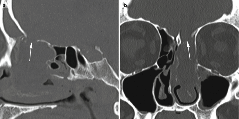
CSF leak. (a) Sagittal CT cisternogram image. (b) Coronal CT cisternogram image. Postoperative changes from prior right ethmoid resection and medial maxillary antrostomy are present. In both images, contrast pooling is detected in one of the ethmoid air cells on the left, immediately below the roof (arrows), consistent with leakage of CSF. A bony defect is not directly visualized

Fig. 73.2
CSF leak. (a) Sagittal CT image without contrast. (b) Coronal CT image without contrast. In both images, a bony defect is seen in the roof of the left ethmoid (arrows), with complete opacification of the left ethmoid air cells and part of the left nasal cavity

Fig. 73.3
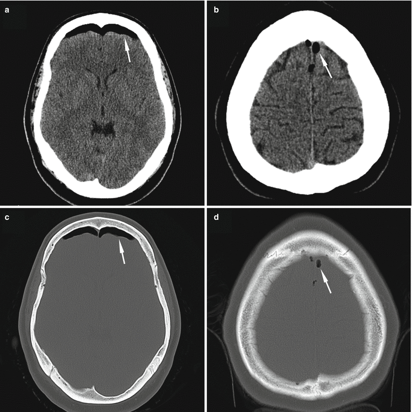
CSF leak. Noncontrast axial CT images in a patient with a postoperative CSF leak following pituitary surgery. Images (a, b) are windowed for brain soft tissue, and images (c, d) are windowed for bone imaging. Pneumocephalus (arrows) can be seen in the frontal region in (a, c) and near the superior sagittal sinus, along the convexity, in (b, d)

Fig. 73.4
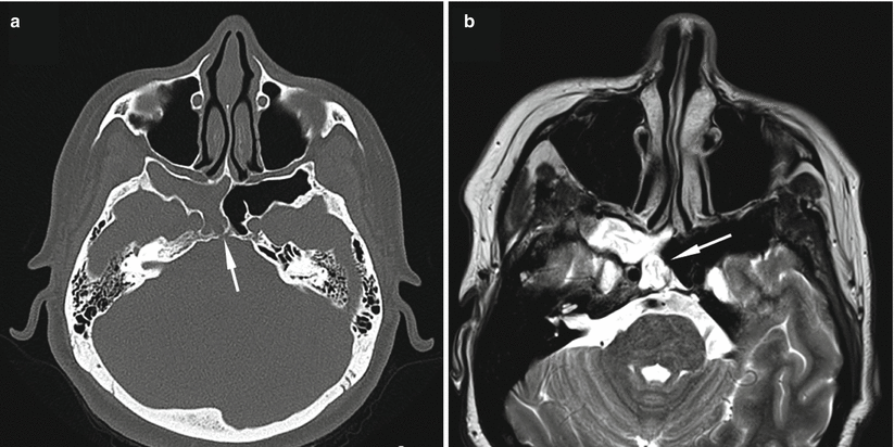
CSF leak. Axial noncontrast CT image (a) and noncontrast T2-weighted MR image (b) in a patient with a CSF leak arising from the clivus and presenting with right-sided rhinorrhea. CT imaging shows the area of bony dehiscence (arrow) and opacification of the right sphenoid sinus (a). MR imaging shows T2-hyperintense CSF pooling (arrow) in the right sphenoid sinus (b)
73.3 Clinical and Surgical Management
73.3.1 Diagnosis of CSF Leaks
In most patients, the diagnosis of CSF rhinorrhea is clinical. The rhinorrhea may be dependent on exertion or on position, worsening when the patient leans forward (“tilt test”). CSF leaks often appear as clear or blood-tinged fluid that looks like a drip from a faucet as a patient tilts forward. Also associated with CSF leaks is the “halo” sign, in which a yellowish ring forms on paper or gauze when CSF is present.
Confirmation of CSF may be done via laboratory analysis for beta-2-transferrin, which is highly specific in differentiating CSF from other clear fluids such as nasal secretions or tears.
A CT scan of the head may show pneumocephalus, which is highly suggestive of a CSF fistula.
In some patients with CSF rhinorrhea, the exact fistula point cannot be identified with standard imaging. In these patients, tests such as CT cisternography or radionuclide cisternography studies can be performed to help identify the site of leakage [18].
Intraoperatively, during a transsphenoidal operation, a Valsalva maneuver should be performed to assess for a CSF leak. Many postoperative CSF leaks develop because the leak was not identified during surgery, rather than representing a failed sellar repair [19].
73.3.2 Management of CSF Rhinorrhea
CSF leaks resolve spontaneously in some patients, depending on the etiology.
Medical management with acetazolamide, which reduces the production of CSF and therefore reduces intracranial pressure, can be used as a temporizing measure. It may cause severe nausea and vomiting, however, which can exacerbate the CSF leak, so acetazolamide should be used only after weighing the risks and benefits.
Lumbar punctures and temporary lumbar drains are often successful treatments for CSF leaks causing rhinorrhea. These maneuvers usually work for smaller leaks and those where there is sufficient tissue in place to form a permanent scar, or seal, at the fistula point.
Many surgical options are available for repair of skull base CSF leaks, including autologous fat and fascia, dural substitutes, collagen sponge, fibrin glue, and dural sealants. Multilayer closures tend to offer the best chance of achieving a permanent repair.
For larger leaks with wide bony deficits, the use of a rigid buttress may be beneficial. This can be crafted from bone (the vomer or turbinate), cartilage, titanium, or an absorbable synthetic plate.
The use of pedicled nasal or temporalis flaps can be highly successful in closing larger skull base defects, greatly adding to the armamentarium of endoscopic skull base surgeons. The most commonly used flap is the pedicled nasoseptal flap [21–23].
Patients with refractory CSF leaks despite multiple attempts at surgical repair often have underlying intracranial hypertension, and the definitive management option may be insertion of a CSF-diverting shunt, such as a permanent ventriculoperitoneal or lumbar-peritoneal shunt.
Intracranial pressure may increase following successful repair of a CSF leak, causing new symptoms or leaks in other locations [4].
Outcomes following CSF leak repair are generally excellent, with the leak being repaired on the first attempt in over 90 % of patients [4, 24].
Stay updated, free articles. Join our Telegram channel

Full access? Get Clinical Tree


