Stroke and Vascular Neurology


Stroke and Vascular Neurology


Kevin A. Shapiro

Ferdinando S. Buonanno


OVERVIEW OF STROKE IN INFANTS AND CHILDREN

DEFINITION: A clinical syndrome of rapidly developing focal or global disturbance of brain function lasting >24 h or leading to death with no obvious nonvascular cause (WHO). Problematic in children because (1) children may have infarcts despite rapid resolution of symptoms; (2) presentation may be nonspecific (seizures, headaches); (3) children often present with stroke mimics: migraine, postictal paresis, ischemia due to metabolic disease (e.g., mitochondrial disorders).1

EPIDEMIOLOGY: Varies by age. Neonates: Estimates vary but rate of stroke is ˜1:4,000 to 1:5,000 live births,2 equivalent to annual incidence of large-vessel ischemic stroke in adults; if late-presenting presumed perinatal strokes are included, incidence may be as high as ˜1:2,500; 80% ischemic, remainder cerebral venous thrombosis (CVT) or hemorrhage. Children: Variable estimates; in children <16 y, most studies estimate incidence of 2.3 to 2.7/100,000 per year; however, some studies suggest incidence as high as 13/100,000 per year. Approximately 55% are ischemic and 45% are hemorrhagic, although the proportion varies across studies; about 10% of hemorrhages result from CSVT. All varieties of childhood stroke have a male predominance; African American children have a higher risk even after accounting for sickle cell disease (SCD).1,3,4

RISK FACTORS: Risk factors for all stroke types include infections, leukocytosis, anemia, prothrombotic state. Arterial ischemic stroke (AIS): Underlying conditions include SCD, heart disease; acquired conditions include head and neck trauma. Venous stroke: Underlying conditions include inflammatory bowel disease, autoimmune disorders; acquired conditions include dehydration, infections of the head and neck (e.g., otitis media).


STROKE AND CEREBROVASCULAR DISEASE IN NEONATES


Perinatal Ischemic Stroke

Defined as stroke occurring between 28 wk gestation and 7 d of life.

PATHOPHYSIOLOGY: Thought to occur most often before ˜72 h of postnatal life, as early as the second trimester; usually involves middle cerebral artery (MCA) territory (L > R); may result from thrombosis of intracranial vessels, cardioembolism, or embolism from extracranial vessels or placenta (through patent foramen ovale [PFO]).

Maternal risk factors: Maternal infertility, oligohydramnios, preeclampsia, prolonged rupture of membranes, umbilical cord abnormality, chorioamnionitis, primiparity, monochorionic twin gestation, maternal drug use (cocaine or amphetamines), diabetes, antiphospholipid antibodies.


Fetal risk factors: Prothrombotic disorder (68%) including: lipoprotein(a) (20%), factor V Leiden, prothrombin gene mutation, MTHFR mutation, antiphospholipid antibodies, and protein C deficiency; polycythemia; congenital heart defects.

Peri- and postnatal risk factors: Traumatic delivery, perinatal asphyxia, dehydration, hypotension, infection, portal vein thrombosis, intravascular catheters.

CLINICAL PRESENTATION: May present in the immediate postnatal period or after several months or years (presumed perinatal ischemic stroke). Early (<72 h): Typically seizures, usually focal motor; hypotonia; apnea; encephalopathy; may also have no clinical signs or symptoms (50%). Late (>4-6 mo): Early handedness, seizures, or developmental delay.5

EVALUATION: Should focus on establishing presence of injury and potential etiologic mechanisms.


  • Radiology: Cranial ultrasound has low sensitivity for detecting AIS; unilateral decreases in MCA flow velocity may be seen. Computed tomography (CT) is insensitive for venous thrombosis and early AIS, but relatively quick and sensitive for hemorrhagic lesions. Magnetic resonance imaging (MRI) is modality of choice for arterial stroke in the newborn; diffusion-weighted imaging (DWI) can reliably detect ischemic injury within 24 h of onset (Fig. 18.1), but may pseudonormalize within 7 d (although at this stage T1- and T2-weighted imaging should be abnormal). Encephalomalacia and calvarial hypertrophy (Dyke-Davidoff-Masson phenomenon) can be seen as late sequelae of perinatal stroke (Fig. 18.2). Magnetic resonance angiography (MRA) of the neck should be obtained if there is a history of traumatic delivery or other reason to suspect neck trauma resulting in injury/dissection of cervicocephalic vessels. Echocardiography should be performed to exclude congenital heart defects and intramural thrombi. Doppler ultrasound of the umbilical vessels and portal vein should be performed if umbilical catheters are present.


  • Laboratory: Initial laboratory evaluation should include hematocrit and prothrombotic risk factors: activated protein C resistance for factor V Leiden, protein C, protein S, lipoprotein(a), antithrombin III, prothrombin gene mutation, homocysteine/MTHFR gene mutation, antiphospholipid antibodies. Repeat testing in 6 to 8 wk is recommended for protein C, protein S, antithrombin III, lipoprotein(a), and antiphospholipid antibodies.6


  • Placental pathology: Often informative due to presence of placental infarcts, vasculopathy, or evidence of perinatal infection.


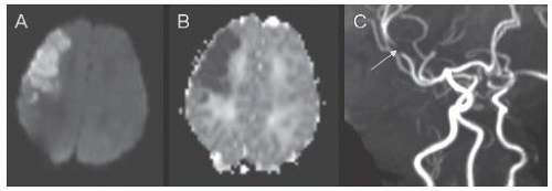
FIGURE 18.1 Corresponding axial slices from diffusion-weighted imaging (DWI) (A) and apparent diffusion coefficient (ADC) (B) MR sequences demonstrating a right frontal arterial ischemic infarction in a 2-day-old boy who developed focal seizures at 29 h of life. The MR angiogram (C) shows focal occlusion of the anterior division of the right middle cerebral artery (arrow).


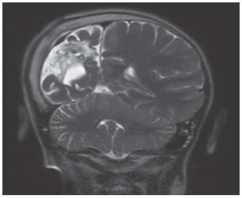
FIGURE 18.2 Coronal T2-Weighted MR Image from an 8-Year-Old Boy with Presumed Perinatal Arterial Ischemic Infarct Affecting the Right Middle Cerebral Artery. The image demonstrates encephalomalacia and atrophy of the right hemisphere, with overlying calvarial hypertrophy (Dyke-Davidoff-Masson phenomenon).

MANAGEMENT: Mainstay of management includes basic neuroprotective strategies: normothermia, normotension, normoglycemia, and control of seizures. Treatment of dehydration and anemia are recommended. Consider folate and B-complex vitamins in infants with MTHFR mutations. Consider anticoagulation in infants with severe thrombophilia or multiple emboli; may also be considered if the etiology is clearly cardioembolic; suggested regimen is enoxaparin 1.5 mg/kg SC b.i.d. Thrombolytic agents are not currently recommended.

OUTCOME: Vast majority (>95%) survive into adulthood,7 but about 64% of children have one or more neurologic disabilities.8,9

Risk factors for disability: Concomitant involvement of cerebral hemisphere, internal capsule, and basal ganglia is almost always associated with abnormal outcome. Involvement of only one or two of these regions may be associated with normal outcome; involvement of internal capsule is most highly predictive of abnormal motor outcome. Presence of neonatal seizures and/or abnormal EEG increases risk of abnormal neurodevelopmental outcome.10,11 Neonatal encephalopathy is predictive of poorer outcomes.12

Types of disability: Hemiplegic cerebral palsy is a common outcome (37% of children with perinatal AIS recognized in newborn period).8 A seizure disorder may develop, though incidence of epilepsy after the perinatal period is <50% in most studies.13 Other neurologic comorbidities include delayed language development, visual abnormalities, cognitive and behavioral disorders.

Recurrence of stroke is rare (<2%), although long-term follow-up has been limited; risk factors for recurrence include prothrombotic states and complex congenital heart disease.14


Neonatal Hemorrhage

For germinal matrix hemorrhage in preterm infants, see Chapter 19.

EPIDEMIOLOGY: Overall prevalence of intracranial hemorrhage may be as high as 26% if preterm intraventricular hemorrhage and mild bleeding in vaginally delivered term neonates are included. Symptomatic perinatal hemorrhage occurs in ˜6/100,000 live births. Types of perinatal hemorrhagic stroke include subdural, subarachnoid, intracerebral, and intraventricular hemorrhage.

RISK FACTORS: Thrombocytopenia; cavernous malformations; genetic diseases (e.g., COL4A1 mutations); maternal consumption of salicylates, anticoagulants, or anticonvulsants. Infants born to mothers taking warfarin, barbiturates, or phenytoin should receive a higher dose of vitamin K after birth to prevent bleeding. Hemorrhage may also be secondary to CVT.

PRESENTATION: Most present acutely with encephalopathy and/or seizures.

DIAGNOSIS: Often discovered by head ultrasound, but this modality lacks sensitivity and specificity; can be confirmed radiographically by CT or MRI (with gradient-echo and susceptibility-weighted imaging). MRA and MRV are indicated to exclude vascular malformations and venous thrombosis. Laboratory evaluation should include platelet count, coagulation studies, screening for bleeding diatheses (e.g., von Willebrand disease); if thrombocytopenia is present, maternal and infant platelet antibodies should be checked.

MANAGEMENT: Correct underlying coagulopathy with platelet transfusion, coagulation factors, and vitamin K is important. Basic neuroprotective strategies include maintaining normothermia, normotension, normoglycemia, and control of seizures. Surgical evacuation of intracranial hematoma is indicated if there is evidence of increased intracranial pressure. Head circumference should be monitored closely in infants at risk for posthemorrhagic hydrocephalus (e.g., IVH); in these cases, ventricular shunting or third ventriculostomy may be required.


Neonatal CVT

EPIDEMIOLOGY: Neonates account for 43% of CVT in childhood.15 Superficial venous system is most often involved. Deep venous thrombosis is a common cause of intraventricular hemorrhage in term neonates.

Risk factors in neonates include infection, dehydration, and anemia.

PRESENTATION: Typically presents with seizures or encephalopathy in the 1st few weeks of life.

DIAGNOSIS: In neonates, hemorrhage associated with venous thrombosis may be demonstrated on head ultrasound; Doppler ultrasound may be used to interrogate flow within the dural sinuses. On MRI, subacute thrombus may be T1 hyperintense; acute thrombus is isointense on T1-weighted images and hypointense on T2-weighted images; T2* (susceptibility)-weighted imaging is most sensitive of MR sequences (Fig. 18.3). MR venography can confirm presence of thrombus.

MANAGEMENT: Predisposing factors such as dehydration and systemic infection should be treated. Low molecular weight heparin may be used in neonates with symptomatic CVT, although there is a risk of ICH.

PROGNOSIS: Developmental delays noted in 28% to 58% of children after neonatal CVT; 6% to 28% have cerebral palsy. Seizures after the neonatal
period occur in 6% to 20%. CVT with infarction is associated with worse neurodevelopmental outcomes than CVT without infarction. Recurrence risk is thought to be low, although data are limited.15,16


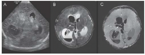
FIGURE 18.3 Coronal transfontanel head ultrasound image (A) from a 1-day-old boy with seizures, showing bilateral intraventricular hemorrhage and associated ventriculomegaly. Axial T2-weighted (B) and susceptibility-weighted (C) MR images illustrate thrombosis of the right internal cerebral vein (arrows).


AIS in Children

Most AIS in children are due to arteriopathy, and vascular imaging is normal in only 21% of children with AIS.17,18 Many arteriopathies are acquired, including cervicocephalic artery dissection, post-varicella arteriopathy, and transient cerebral arteriopathy of childhood; others include arteriopathy due to moyamoya syndrome and SCD. Nonarteriopathic ischemic strokes may be due to cardiac disease or thrombophilia without obvious arteriopathy. Specific causes are discussed in detail below.

RISK FACTORS: In 2/3 of cases, a vascular risk factor can be identified. Risk of recurrence is 6% to 14%; predictors include elevated lipoprotein(a), protein C deficiency, other thrombophilic states, and moyamoya syndrome.19,20 Substances associated with increased risk of ischemic stroke (generally in adults) include cocaine, amphetamines, ecstasy, phentermine, ephedrine/pseudoephedrine.

Hypercoagulable states: One or more prothrombotic states can be identified in 20% to 50% of children presenting with AIS.21

PRESENTATION: Acute onset of a new neurologic deficit including hemiparesis, aphasia, visual disturbance or at times, seizure. Most children with nonarteriopathic causes of AIS have abrupt onset (72%); children with arteriopathic causes of stroke more often have a progressive, stuttering, or recurring onset (68%).22

EVALUATION: See Table 18.1.

Radiology: MRI is modality of choice for evaluation of suspected stroke; should include T1, T2/FLAIR, T2* (susceptibility), DWI with ADC. CT is sensitive for detection of hemorrhage and may identify early findings consistent with ischemic stroke; rapid acquisition useful in unstable patients. Vascular imaging should be obtained in all children with stroke; MRA is typically of high quality in children; CTA has high spatial resolution but results in a large radiation dose. Catheter angiography is preferred for evaluation of tertiary branches and small cerebral arteries (e.g., in suspected vasculitis and moyamoya); potential complications include damage to vessels at sites of access, catheter-induced dissection or perforation, thrombosis, and complications of contrast administration or anesthesia.


TABLE 18.1 Recommended Evaluation for Children with Arterial Ischemic Stroke


Radiology


MRI with contrast-enhanced MRA of head and neck


  • Consider T1 fat-saturated images of neck if high suspicion for arterial dissection


  • CT and CTA may be helpful if MRI/MRA contraindicated


Echocardiogram


Transthoracic echocardiogram (TTE) with agitated saline to evaluate for:


  • structural heart disease


  • cardiomyopathy


  • mural thrombus


  • intracardiac or extracardiac shunts


Laboratory


  • CBC with differential, electrolytes, BUN/Cr


  • PT, PTT, fibrinogen, d-dimer


  • Urine β-HCG (in postpubertal girls)


  • Serum and urine toxicology screen


  • Blood cultures


  • Protein C, protein S, antithrombin III, activated protein C resistance (factor V Leiden), prothrombin gene mutation


  • Homocysteine


  • ESR, CRP, ANA, anticardiolipin antibodies, anti-β 2 glycoprotein antibodies, lupus anticoagulant


  • Lipoprotein panel


  • Hemoglobin electrophoresis


MANAGEMENT: Depends on underlying etiology.


  • Supportive measures include avoidance of hypoxemia and hyperthermia, maintaining normal blood glucose. Thrombolytic therapy (tPA) for acute stroke is currently not recommended in children due to lack of evidence, but is sometimes used in older adolescents who meet adult tPA eligibility criteria.


  • Short-term anticoagulation may be initiated in children until the cause of stroke is determined and continued for up to 1 wk. Low molecular weight heparin: 1 mg/kg every 12 h; monitor anti-factor Xa activity drawn 4 to 6 h after dose (goal 0.5-1 U/mL). Unfractionated heparin may also be used, especially in situations where need for reversal of anticoagulation is anticipated.


  • Long-term anticoagulation is indicated in children with risk of recurrent cardiac embolism, arterial dissection, and certain hypercoagulable states. Low molecular weight heparin usually initial choice; may be continued or transitioned to warfarin or newer oral anticoagulants (e.g., rivaroxaban).


  • Antiplatelet agents may be used for secondary prevention of strokes in children without SCD who are not known to have high recurrent risk of embolism or severe hypercoagulability. Aspirin is usually dosed at 3 to 5 mg/kg/d; if dose-related side effects occur, may reduce to 1 to 3 mg/kg/d. Clopidogrel 1 mg/kg/d has been used in children unable to tolerate aspirin.


  • If homocysteine is elevated, it is reasonable to supplement with folate, B12, or pyridoxine. Oral contraceptives should be discontinued in children with ischemic stroke.

PROGNOSIS: Mortality in childhood ischemic stroke may reach 20%, about half of which is related to underlying illness rather than stroke itself.
Between 50% and 80% of surviving children have neurologic sequelae, most commonly hemiparesis; other problems include poor attention, behavioral problems, and cognitive deficits. Poorer outcome is associated with systemic disease, multiple risk factors, infarct size, cortical involvement, thromboembolism, and moyamoya.1


NONATHEROSCLEROTIC VASCULOPATHIES


Cervicocephalic Artery Dissection

EPIDEMIOLOGY: Carotid and vertebral artery dissection accounts for ˜7.5% of childhood ischemic stroke23,24; may occur spontaneously or after blunt or penetrating trauma. More common in younger patients, patients with family history of dissection; associated with connective tissue disorders (Ehlers-Danlos type IV, Marfan syndrome), aortic coarctation, autosomal dominant polycystic kidney disease, osteogenesis imperfecta, atherosclerosis, extreme arterial tortuosity, moyamoya syndrome, and pharyngeal infections.

DIAGNOSIS: Angiography (conventional, MRA, CTA) may demonstrate “string sign”; double lumen sign; short, smooth, tapered stenosis; occlusion of parent artery. MRI with fat-saturated T1 imaging of the neck and contrast-enhanced MRA are generally used as first-line evaluation; CTA not recommended in children.

MANAGEMENT: Studies in adults show no evidence for superiority of antiplatelet therapy or anticoagulation; either may be used in children. Anticoagulation with LMWH, warfarin (goal INR 2.0-3.0), or newer oral anticoagulants (e.g., rivaroxaban) is typically continued for 3 to 6 mo, but may be extended if symptoms recur; anticoagulation is avoided in patients with intracranial dissections (due to risk of subarachnoid hemorrhage) and at least initially in patients with large infarcts (due to risk of hemorrhagic conversion). Antiplatelet therapy is a reasonable alternative and may be continued longer than 6 mo, especially when radiologic abnormalities persist.


Moyamoya Disease and Moyamoya Syndrome

Chronic progressive stenosis of intracranial internal carotid arteries; less often stenosis of middle, anterior, and posterior cerebral arteries and basilar artery; may be unilateral or bilateral; moyamoya (Japanese: “puff of smoke”) refers to hazy angiographic appearance of fine collateral vessels. Accounts for up to 6% of all childhood strokes.25,26


EPIDEMIOLOGY

Moyamoya disease: Idiopathic; most common in Japan (3 per 100,000); rarer in the United States (0.086 per 100,000); more frequent in Asian (OR 4.6) and black children (OR 2.2) compared to white children.27,28 Bimodal age distribution: children in the 1st decade of life (more likely to present with ischemic events) and adults in the 4th decade (more likely to present with hemorrhage). Genetic factors likely important (familial incidence of affected first-degree relatives is 6%-12%), but poorly understood; has been associated with specific HLA haplotypes (HLA B40, B52).

Moyamoya syndrome: Diagnosed in individuals with well-recognized associated conditions, including history of cranial radiation, Down syndrome, neurofibromatosis 1, SCD.

PRESENTATION: Strokes and transient ischemic symptoms classically occur in the setting of hyperventilation and crying (due to cerebral vasoconstriction), coughing or straining (due to decreased cerebral perfusion pressure), or fever (due to increased metabolic demand).


DIAGNOSIS: Based on distinct angiographic appearance on MRA, CTA, or conventional angiography, but other modalities may yield important diagnostic information.


  • CT/MRI may show small areas of infarction in cortical watershed zones, basal ganglia, deep white matter, or periventricular regions. MRI may show absence of flow voids in ICA, MCA, and ACA with abnormal flow voids from basal ganglia and thalamic collaterals (Fig. 18.4).


  • EEG shows slowing of the background rhythm after hyperventilation (“re-buildup”).


  • TCD may assist with diagnosis and postoperative follow-up.


  • Various techniques used to determine resting perfusion/blood flow reserve and to predict improvement in functional perfusion after therapy (xenon CT, PET, MR perfusion, acetazolamide SPECT).


  • Screening (with MRA) may be considered in individuals with common high risk-associated conditions or prior history of unilateral disease (27% eventually develop bilateral involvement).

MANAGEMENT: Despite extensive literature, there are no controlled clinical trials to guide medical vs. surgical therapy or type of surgical therapy.


  • Surgical revascularization (by direct or indirect anastomoses) is used in patients with cognitive decline or recurrent or progressive symptoms.25 Meta-analyses suggest that 87% derive symptomatic benefit from revascularization, but no differences between types of procedures.29 Potential complications: postoperative stroke, spontaneous or traumatic subdural hematoma, ICH. Perioperative management includes sedation and analgesia (to prevent crying and hyperventilation), avoidance of hypotension, hypovolemia, hyperthermia, and hypocarbia.


  • Antiplatelet therapy (aspirin 3-5 mg/kg) may be used when patient has a poor operative risk or has mild disease, or routinely after surgery.


  • Calcium channel blockers may improve intractable headaches and reduce frequency and severity of refractory TIAs.


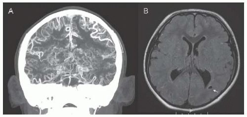
FIGURE 18.4 CTA (A) and FLAIR MRI (B) in an 8-year-old boy with trisomy 21 and moyamoya syndrome. CTA shows terminal stenosis of the bilateral internal carotid arteries distal to the origin of the posterior cerebral arteries, with exuberant deep collaterals (moyamoya vessels) as well as pial collaterals. MRI shows chronic white matter infarcts (arrow) in the posterior-middle cerebral artery border zones.


Fibromuscular Dysplasia

Nonatherosclerotic segmental noninflammatory arteriopathy, typically affecting renal arteries and extracranial ICA (3-4 cm from bifurcation); 20% to 30% have cerebrovascular involvement. Stroke due to stenosis or dissection of affected artery, or embolism from stenotic artery; aneurysms in ˜7% of patients.1 Antiplatelet therapy or anticoagulants often used; angioplasty and/or stenting or endarterectomy can be considered if symptoms recur. Other surgical therapies include coiling of cerebral aneurysms.


Fabry Disease

X-linked sphingolipidosis caused by deficiency of α-galactosidase, leading to accumulation of glycosphingolipids with α-D-galactosyl moieties. Primary cause of vasculopathy is progressive accumulation of globotriaosylceramide (Gb3) in endothelial and vascular smooth muscle cells, causing small-vessel stenosis/occlusion, more prominent in the vertebrobasilar system. Additional factors leading to increased stroke risk may include higher prevalence of cardiac disease and BP dysregulation (hyper- or hypotension), dilation and dolichoectasia of large vessels leading to flow stagnation and thrombosis, prothrombotic state, premature atherosclerosis, and autoimmunity.30

EPIDEMIOLOGY: Incidence ˜1/117,000 live births and ˜1/40,000 men31; >400 mutations in α-GAL gene have been identified, including de novo mutations. Estimated that 1.2% of cryptogenic strokes in patients younger than 55 y are attributable to mutations in α-GAL.32 Stroke more common in women with FD (27%) than men (21%), but women tend to be older at onset of stroke (43.4 y) compared to men (28.8 y).33,34

CLINICAL PRESENTATION: Stroke may be the first manifestation of Fabry disease34 and commonly affects the posterior circulation; brain MRI typically shows progressive white matter T2 hyperintensities characteristic of small-vessel disease. Manifestations outside the CNS in children include pain crises due to peripheral neuropathy (acroparesthesias), fever, hypohidrosis, and exercise intolerance; numerous organ-specific manifestations (Table 18.2).


TABLE 18.2 Nonneurologic Manifestations of Fabry Disease


Only gold members can continue reading. Log In or Register to continue

Stay updated, free articles. Join our Telegram channel

Jun 20, 2016 | Posted by in NEUROLOGY | Comments Off on Stroke and Vascular Neurology

Full access? Get Clinical Tree

Get Clinical Tree app for offline access

Dermatologic


Angiokeratoma


Hypohidrosis/anhidrosis


Renal