Sudden Death of Central Nervous System Origin in Adults


Sudden Death of Central Nervous System Origin in Adults


Ana Rubio


Sudden death constitutes one of the most frequent scenarios in the routine forensic pathology practice, and the neuropathologist may be consulted in such cases to rule out brain pathology as causative of death. Sudden death may be defined as either death without warning, or as death occurring shortly after presentation of symptoms. How shortly after initiation of symptoms death must occur to be considered sudden ranges from less than 1 hour1, 2, 3 to less than 1 day.4, 5, 6, 7 Some deaths are truly instantaneous, as seen in individuals with a witnessed collapse. Not all sudden deaths are unexpected; people with terminal illnesses may die suddenly, although not unexpectedly, and as such, they are not the object of this chapter. Figure 14.1 depicts the procedures we follow when working up cases of sudden death in forensic pathology. In our experience, most cases fit one of the following situations: the person is found dead, death is witnessed, or the individual dies either during transportation to the emergency room or at the hospital before a diagnosis is reached. Investigation of the circumstances surrounding death, knowledge of the clinical history, examination of the body, and performance of toxicologic analyses help to rule out trauma or intoxication as factors contributing to or causative of death; in cases in which such factors are present, the manner of death is not natural. Determining the cause of natural, sudden, unexpected death may be challenging and alternatively rewarding or frustrating. In approximately 5% of cases, the cause of death remains unknown even after a complete investigation and thorough postmortem examination, 8, 9 and depending on the circumstances, the cause of death may be listed as probable cardiac arrhythmia or as undetermined.


HEART OR BRAIN

Overall, the majority of natural, sudden, unexpected deaths studied in medical examiner offices are of cardiac origin, 10, 11, 12 and central nervous system (CNS) pathology is the cause of death in less than one-fifth of sudden deaths. The physiologic and physiopathologic connections between heart and brain have important clinical and pathologic implications in the evaluation of sudden death. It is not uncommon for sudden cardiac deaths to present with seizure-like movements preceding terminal collapse. These tonic-clonic-like episodes are secondary to the cerebral hypoxia induced by the ineffective heart function. The clinical differentiation between syncope and idiopathic seizures may be so challenging, that the phrase “when it looks like brain—think of heart!” has been uttered.13 It is also our experience (unpublished review) that a significant percentage of witnessed sudden cardiac deaths have seizure-like episodes as the main sign immediately preceding death. Alternatively, deaths caused by brain pathology may manifest with cardiac signs. One of the mechanisms of death in epilepsy is cardiac arrhythmia during seizures or in the postictal period, and a variety of arrhythmias have been recorded in individuals monitored during or immediately after seizures14, 15 and other situations of sudden death resulting from CNS pathology, such as head trauma, 16 subarachnoid hemorrhage, 17 and neurosurgical manipulation of the brain, 18 suggesting that the mechanism of death in a percentage of deaths of CNS-related cause is cardiogenically mediated.


EPIDEMIOLOGY

The best-understood mechanism of CNS-related sudden death is acute elevation of intracranial pressure (resulting from intracranial hemorrhage, brain swelling, or acute blockage of cerebrospinal fluid [CSF] circulation, among other causes), but localized pathology in vulnerable topographic areas of the brain can also be the culprit. Occasionally, sudden death results from acute decompensation of previously undiagnosed intracranial pathology (such as hemorrhage expanding a tumor). An 8-year review of intracranial lesions responsible for sudden death in Scotland identified 218 such cases, 19 of which 60% were due to sudden unexplained death in epilepsy, 37% to intracranial hemorrhage, 2% to meningitis, and 0.5% to brain tumors. In our experience at the Office of the Chief Medical Examiner of the State of Maryland, given the inclusion criteria for cases to reach our office (deaths resulting from violence or unexpected/unwitnessed deaths), the majority of adult deaths certified as natural fit criteria for sudden death. A sizable percentage of these deaths occur in individuals with chronic illnesses, even though they may not be expected to die. A 10-year review of natural deaths in adults in the state of Maryland from 1997 to 2006 showed that of 48, 878 individuals studied at the office, in 2, 004 cases (4.1%), the primary cause of death was CNS pathology (Tables 14.1 and 14.2). Below, we review the most common diagnostic groups responsible for sudden unexpected death caused by CNS pathology.


SUDDEN UNEXPLAINED DEATH IN EPILEPSY

Total mortality among epileptics is two to three times higher than it is in people without seizures, 20 and the risk for sudden death is 24 times greater in epileptics.21 This higher mortality is not only due to natural causes, but accidents, such as drowning, motor
vehicle, or industrial mishaps, which are more common among epileptics than in the general population.22, 23 The classical scenario in accidental deaths in epilepsy is when an individual with a history of seizures is witnessed to have a seizure, and the resultant erratic driving causes an accident. At other times, epileptics may drown, and the logical inference is that they suffered a seizure that resulted in drowning. Further, seizures causing an accident may be the most likely explanation for death in cases with or without a history of seizures, when a seizure focus is identified at autopsy and no other cause of death or any other explanation for the accident is apparent, as in Case 14.1.


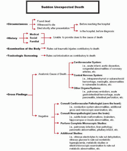
FIG. 14.1. Schematic representation of the algorithm followed in the work-up of sudden deaths at the Office of the Chief Medical Examiner of the State of Maryland.

The relevance of sudden death in epilepsy has been recognized for some time among the forensic and neuropathologic communities, but a long-lasting myth among clinicians, now debunked, maintained that seizures per se did not have a fatal consequence. In 1954, a letter to the Journal of the American Medical Association regarding mortality in epilepsy expressed that “pathologists generally have felt that the only possible cause for death in the course of epilepsy is the condition known as status epilepticus. It is often said that no one dies in a seizure. From the pathologist’s standpoint, a seizure is not in itself a cause for death.”24 Much has changed in the last half century since the cited article was written, and forensic pathologists and clinicians alike have learned, by experience acquired through individual cases and through evidence resulting from controlled studies, that seizures themselves can cause death.25, 26 A significant proportion of epileptic deaths that fit sudden death criteria have a negative postmortem examination (gross and microscopic studies do not elucidate the cause of death), and the cause of death is certified as sudden unexplained death in epilepsy (SUDEP). SUDEP represents 18% of all deaths in epileptics, 27 and the proportion may be as high as 24% to 67% when only chronic epileptics are included in the analysis.25 The incidence of SUDEP among epileptics ranges from 0.35 to 9.3/1, 000 patient-years of follow-up, 21, 28, 29 depending on the characteristics of the individuals included in the assessment. The risk is lower in population-based studies, higher in neurologist-referral groups and clinical trial populations, and highest among groups of epilepsy-related surgical referrals or among subjects with refractory epilepsy.30

SUDEP is defined as the “sudden unexpected, witnessed or unwitnessed, nontraumatic and nondrowning death in patients with epilepsy, with or without evidence of a seizure, and excluding documented status epilepticus, where necropsy does not reveal a toxicologic or anatomical cause of death.”31, 32 Cases fulfilling all the requirements are classified as definite SUDEP; if the results of postmortem examination are not available (an autopsy is not
done, the autopsy results are not complete, or the autopsy is suboptimal because the body is decomposed), cases may be categorized as probable SUDEP.


Significant evidence supports the fact that in the majority of SUDEP cases, death occurs during or after a seizure. Most individuals are found dead in bed, and scene and autopsy findings suggest a recent seizure, such as an awkward body position, ruffled bedding, a bitten tongue or lips, or urinary incontinence20 (Fig. 14.2, e-Figs. 14.2 and 14.3). Another frequent finding is pulmonary edema manifested as a foam cone (e-Fig. 14.4) and/or heavy, wet lungs identified at autopsy. Night supervision (by direct observation or listening devices) may protect against SUDEP, 20, 33 suggesting that close assistance during or after a seizure can be preventive of fatal complications. In witnessed deaths, the majority of cases are seizure related as well. In a series of 15 cases of witnessed death in epileptics, 34 12 deaths occurred during convulsions, one during the aura, one in the postictal period, and one 5 minutes after generalized seizures.


Risk Factors

Risk factors for SUDEP have been detected by observational, 35, 36 case-control, 20, 25, 26, 33, 37 and cohort28 studies, aided by metaanalyses.38, 39 Death in most SUDEP cases occurs in bed, and there is evidence of a terminal seizure. The majority of the studies found that male gender, young age, and severe seizure history were strong predictors of SUDEP. Different studies identified different severity-of-seizures markers associated with SUDEP, such as early age of onset, long seizure history, presence of generalized seizures, poor seizure control, poor compliance with medication, high number or high frequency of seizures, large number of antiepileptic drugs in use, or frequent change of antiepileptic medications. Other predictors include mental and physical stress, developmental or mental retardation, structural brain lesions, and the use of psychotropic drugs.28 Risk factors identified in some studies but not confirmed in others include treatment with specific drugs such as carbamazepine, and excessive alcohol intake.25, 37, 39, 40, 41


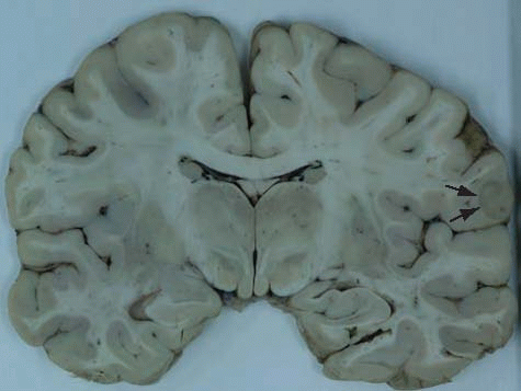
TABLE 14.1 Age and Sex Comparison of Adult Individuals with Natural Death from All Causes and Those with CNS-Related Natural Death: 10-Year Review of Cases Studied at the Office of the Chief Medical Examiner of the State of Maryland (1997-2006)


All Natural Deaths (N = 49, 396)


CNS-Related Deaths (N = 2, 004)


Age, years


Mean (SD)


64.8 (16.4)


56.5 (18.6)


Median


66


53


Range


21-115


21-103


Sex, n (%)


Female


20, 237 (41.5)


897 (44.5)


Male


29, 142 (58.5)


1, 107 (54.5)


Natural deaths of CNS-related pathogenesis represented slightly more than 4% of all adult natural deaths; they occurred at a younger age (≤13 years), and they had a slightly higher proportion of women versus men (44.5% versus 41.5%) than the overall group of all natural deaths.


Mechanism of Death in SUDEP

A variety of mechanisms have been postulated to explain sudden death in epilepsy, including cardiovascular, pulmonary, metabolic, and neurogenic mechanisms.40 Long-term treated epileptics, especially those suffering repeated episodes of status epilepticus, have an increased prevalence of cardiac pathology42 responsible for some cases of sudden death. Arrhythmias reported in epilepsy include prolonged QT interval, ST depression, T-wave inversion, ventricular fibrillation, asystole, bradyarrhythmias, atrial fibrillation, atrial and ventricular premature defibrillation, and sinus and supraventricular tachycardia.43, 44, 45 There are rare case reports of electrocardiographic recordings during seizures. A 51-year-old woman with posttraumatic epilepsy had two episodes of asystole triggered by seizures. The events started in the left hippocampus and progressed to the right hippocampus in 30 seconds; the patient developed myoclonic twitching of the limbs, and approximately 20 seconds later, the electrocardiogram showed asystole lasting 21 and 28 seconds.46 Cardiac arrhythmia may result from ischemia, electrolyte disturbances, arrhythmogenic effect of medications, or activation of the autonomic nervous system and excitation of the heart. Leestma et al.41 found higher heart weight in SUDEP than in controls (using height instead of weight to normalize the heart weight), but these results were not corroborated by Davis and McGwin.47 A case-control study in Denmark48 found a trend toward more fibrosis in the deep and subendocardial
myocardium of SUDEP individuals than in controls. Vagal nerve stimulation procedure has been reported to produce a 40% reduction in seizures27, which may decrease the incidence of SUDEP in cases of intractable epilepsy. Pulmonary insufficiency in SUDEP can result from central apnea, obstructive compromise of the airways, or positional asphyxia related to the seizure episode itself.40 Experimental models of SUDEP have shown a decrease in blood oxygenation, hypercapnia, and metabolic acidosis in the post-ictal period.49 Pulmonary edema is a common finding in SUDEP and, on the basis of animal studies, 49 may be a sign of autonomic dysfunction.


TABLE 14.2 Cause of Death Distribution in Adult Natural Deaths of CNS-Related Pathogenesis


Age


Sex


No.


Mean


SD


Median


Range


Female (%)


Male (%)


Intracranial hemorrhage


755


55.7


15.6


52


21-96


347 (46.0)


408 (54.0)


Seizures


438


44.1


13.8


43


21-98


153 (34.9)


285 (65.1)


Neurodegenerative disorders


261


77.9


12.8


80


31-102


151 (57.8)


110 (42.2)


Hypoxic-ischemic lesions and strokes


244


66.2


18.2


68


23-103


118 (48.4)


126 (51.6)


Intracranial tumors


98


59.2


15.8


60.0


23-91


44 (44.9)


54 (55.1)


Primarily CNS infections


66


41.4


10.0


41


21-63


19 (28.8)


47 (71.2)


Multiple sclerosis


43


53.0


15.0


52


25-88


23 (53.5)


20 (46.5)


Developmental disorder


38


38.1


14.4


33


21-77


12 (31.6)


26 (68.4)


More than half of the cases were due to intracranial hemorrhage. The second most common cause was seizures. Male gender was more common than female gender in all groups, except for the group of deaths associated with neurodegenerative disorders (including dementing illnesses), which were most common among women. The remaining 61 cases (see Table 14.1) were a miscellaneous diagnostic group.


Neuropathology

Examination of SUDEP brains shows abnormalities in 50% to 70% of cases.36, 50, 51, 52, 53, 54 The frequency and type of lesions depend on the cause of seizures (whether traumatic, postinfectious, hypoxic, metabolic, neurodevelopmental, degenerative, or idiopathic), disease duration, and type of therapy. Gross and microscopic abnormalities include lesions causative of seizures, as well as abnormalities resulting from the seizures or seizure therapy. That brain lesions may be either the cause or the result of seizures or seizure treatment is supported by premortem neuroradiologic data and by experimental studies. Serial magnetic resonance imaging (MRI) studies have shown progressive hippocampal atrophy in epileptics corresponding to disease duration.55 A comparative study of subjects with chronic epilepsy, individuals with recent epileptic onset, and normal controls56 has shown generalized neocortical atrophy in the long-term epileptics when compared with the other two groups. Experimental status epilepticus produces brain changes simulating those of hippocampal sclerosis (neuronal loss in CA4 and CA1 layers of Ammon horn).57 Early neuronal death as indicated by positive HSP-70 and c-Jun immuno staining of neurons has been detected in the hippocampus of patients with SUDEP.58 Table 14.3 summarizes the gross and microscopic findings in SUDEP brains (Figs. 14.3, 14.4, 14.5, 14.6 and 14.7, e-Fig. 14.5). The frequency of lesions identified depends on the method of study (fresh versus fixed-brain examination), the expertise of the examiner (nonpathologist versus general or forensic pathologist, versus neurologist or neuropathologist), and the techniques used (e.g., immunohistochemistry, special stains, or other adjuvant methods). As an example, in the Black and Graham study, 19 gross abnormalities were identified in 66% of brains studied in the fixed state, but in only 10% of those cut fresh, suggesting that subtle lesions may be missed when brains are sectioned before fixation. A study of the neuropathologic findings in SUDEP compared with those of a cohort of patients with epilepsy dying of other causes revealed no difference in the type of pathology or the frequency of pathologic findings.25


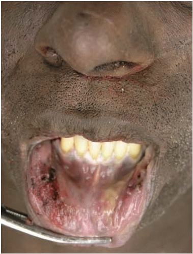
FIG. 14.2. Extensive injuries were observed in the lower retrolabial mucosa of this 52-year-old man with history of a seizure disorder.


INTRACRANIAL HEMORRHAGE

In the following paragraphs, we discuss intracranial hemorrhages as responsible for sudden death, grouped by the main compartment containing the bleeding (see also Chapter 10 for a focused review of vascular pathology). Often, the hemorrhage is not limited to a single anatomic space, and blood may cross into different compartments. For example, subarachnoid hemorrhage may be
the primary location of hemorrhage in ruptured berry aneurysms, but the blood sometimes dissects through the brain parenchyma and accumulates into the ventricular system or perforates the leptomeninges, producing a secondary subdural hematoma. Because the majority of extradural hematomas are traumatic in origin, we exclude them from the current discussion and limit this review to nontraumatic subarachnoid, subdural, intraparenchymal, and intraventricular hemorrhages, which are capable of producing sudden unexpected death in adults.


TABLE 14.3 Neuropathologic Findings in Sudden Unexplained Death in Epilepsy


Gross Examination


Microscopic Studies


General findings


  • Low brain weight


  • Ventriculomegaly


  • Malformations/maldevelopmental lesions


  • Neuronal loss


  • Subpial gliosis


Localized findings


  • Cortical lesions (scars from trauma, ischemia, or infection)


  • Gray-white junction lesions (tumors, abscesses, vascular malformations)


  • Hippocampal atrophy (unilateral or bilateral)


  • Gray matter heterotopia


  • Cerebellar atrophy


  • Ammon horn sclerosis


  • Amygdala sclerosis


  • Neuronal heterotopias


  • Neuronal clusters


  • Oligodendroglial clusters


  • Microdysgenesis


  • Cystic lesions


  • Glial scars


Subarachnoid Hemorrhage

Subarachnoid hemorrhage (SAH) is the most common gross anatomic finding of CNS-related sudden, unexpected, natural death in adults (see Fig. 10.1, e-Fig. 14.7). Approximately 85% of these SAHs are caused by ruptured berry aneurysms59 (e-Figs. 14.8 and 14.9), and the remaining 15% are due to miscellaneous causes described later. Often, the volume of the hemorrhage is not sufficient to explain death, and several other mechanisms have been postulated to explain the demise, such as brain ischemia produced by the vasospasm resulting from a rapid increase in intracranial pressure60, 61 or the irritating effect of blood in the subarachnoid space. Other authors suggest that neurogenic pulmonary edema may result from a vagal reaction induced by diencephalic or brainstem compression.62, 63 Finally, a fatal cardiac arrhythmia64, 65 with cardiac arrest66 may result from a reflex-like effect initiated by the hemorrhage.


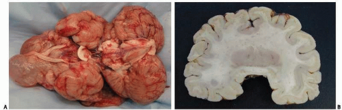
FIG. 14.3. Seizures are a common clinical component of acquired (A) or congenital brain lesions (B). A shows the basilar aspect of the brain of a 16-year-old girl with cerebral palsy and seizures. The brain weighed 280 g and had severe encephalomalacia of the cerebral cortex and white matter. B displays a coronal section of the brain of a 16-year-old girl with the clinical diagnosis of cerebral palsy. Examination of the 720-g brain showed small frontal lobes; shallow, incomplete, interhemispheric fissure; and absence of optic and olfactory nerves (Alobar holoprosencephaly).


Ruptured Berry Aneurysms

In an estimated 12% of the cases, ruptured berry aneurysms manifest as sudden death, 67, 68 and in approximately 36% of these cases, death is witnessed.69 Ruptured berry aneurysms account for 1.5% of all natural deaths studied in a medical examiner office, 69 and in approximately half of the cases, the affected individuals lacked any previous complaints. Aneurysms are most often located in the anterior circulation (branching points of anterior and middle cerebral arteries and proximal branches), but the exact distribution varies, depending on the methodology and type of individuals studied. When only medical examiner cases of sudden death are included in epidemiologic analyses, the representation of aneurysms affecting the posterior circulation is higher than when the group studied includes cases admitted to the hospital after aneurysmal rupture, cases of intact aneurysms incidentally found in radiologic studies performed for unrelated reasons, or unruptured aneurysms incidentally found in autopsy series (Fig. 14.8). This difference in frequency of distribution suggests that posterior circulation aneurysms are either more likely to rupture, that once they rupture they are more lethal, or both. A population-based study67 comparing the aneurysmal location in patients with
ruptured berry aneurysms presenting with sudden death and those that arrived alive to the hospital found that 38% of the aneurysms involved the posterior circulation in the first group and 14% in the second. A meta-analysis showed that the risk of sudden death in ruptured vertebrobasilar aneurysms was more than twice the risk for death in ruptured anterior circulation aneurysms.68, 70 Risk factors associated with aneurysmal rupture include female gender, middle age, smoking, and history of hypertension (albeit undiagnosed or untreated).

Only gold members can continue reading. Log In or Register to continue

Stay updated, free articles. Join our Telegram channel

Aug 1, 2016 | Posted by in NEUROLOGY | Comments Off on Sudden Death of Central Nervous System Origin in Adults

Full access? Get Clinical Tree

Get Clinical Tree app for offline access