(1)
Hand Surgery Department of Clinical Sciences, Malmö Lund University Skäne University Hospital, Malmö, Sweden
Abstract
The exceptionally well-developed sensory functions of the hand are based on a delicate interaction between hand and brain. Tactile stimuli on the hand activate sensory receptors that are responsive to pressure, vibration, tension, cold, heat and nociceptive stimuli. Sensory signals reach the somatosensory cortex in the contralateral hemisphere via nerve pathways in the spinal cord. A cortical body map was defined more than 75 years ago showing that all parts of the body are represented within specific areas in somatosensory and motor cortices. The hand, face, mouth and tongue occupy the major parts, reflecting their delicate sensory and motor functions. Processing of tactile stimuli from the hand is an extremely complicated physiological process based on several parallel body maps processing information from various types of receptors in the skin as well as deeper tissue structures.
What are the secrets behind the refined motor and sensory properties of our hands? How can the sensibility in our hands be so well developed, and how can the hand and the brain interact so perfectly when we perform delicate precision tasks and when we perceive and recognise the roughness in the surface of a brick, the softness of silk and velvet or the shape and structure of a walnut?
When we touch an object or move our fingers over an irregular surface, small displacements of the skin of the fingertips create vibrations. In the hand, and especially in the hairless skin of the finger pulps, there are large numbers of sensory receptors (mechanoreceptors), responding to touch (pressure), vibrations or stretching (Fig. 8.1) [1–8]. There are also various types of free nerve endings that are activated by cold, heat or painful stimuli (nociceptive stimuli). Tactile stimulation of the sensory receptors of the fingers generates electric signals in large myelinated nerve fibres of the hand, and via nerve trunks in the hand and arm and pathways in the spinal cord, these sensory signals reach the somatosensory cortex, primarily in the contralateral hemisphere in the brain (Fig. 8.2). Thus, the sensory stimuli occur in the fingertips, but the perception and sensory experiences occur in the brain. We are dealing with an extremely complicated system of nervous pathways and relay stations in the brainstem and in the thalamus before the signals reach the somatosensory cortex; much can happen along the route from the hand to the brain that might influence the final sensory experience [2].
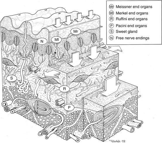
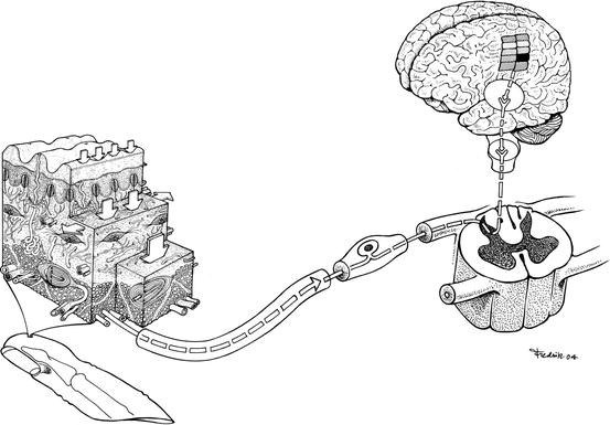
Fig. 8.1
Superficial skin segment from the pulp of a finger with the ridges of the fingerprint visible at the top. Mechanoreceptors in the skin of the hand and fingers detect pressure (Merkel end organs), vibration (Meissner and Pacini end organs) and tension and stretching (Ruffini end organs) (Illustration: Fredrik Johansson. From Lundborg [2])
Fig. 8.2
The sensory pathway of sensory impulses induced by tactile stimulation of a finger. Afferent signals from cutaneous mechanoreceptors travel through large myelinated nerve fibres surrounded by a myelin sheath. They reach the somatosensory cortex after passing the dorsal root ganglia, up the dorsal column of the spinal cord via the medial meniscus pathway and the intermediate relay stations in the cuneate nucleus in the brainstem and the ventroposterior nucleus of the thalamus. Signals, elicited by touch, are primarily transmitted to the contralateral hemisphere
The sensory mechanoreceptors in the hand interact to send appropriate sensory information to the brain when the hand is touched or when the hand is involved in an active ‘act of touch’ process where the sensory feedback is essential – for instance when we make meatballs, knead dough, test the consistency of bread or move our fingers across a surface to feel its texture. The sensory receptors that react to vibrations (Pacini end organs and Meissner end organs) are located in deep and superficial layers and are activated by vibrations within large and small cutaneous areas, respectively (Fig. 8.1). Merkel end organs adapt slowly and mainly respond to vibrations below 50 Hz, while Pacini end organs, which are fast adapting, mainly react to vibrations above 50 Hz. Merkel end organs, also called the Merkel disc receptors, located superficially at the centre of the papillary ridges of the skin, adapt slowly and react to static pressure; Ruffini end organs, found in deep dermal layers primarily on the dorsal aspect of the hand, are activated by tension and stretching of the skin [1–4]. Ruffini end organs in the skin on the back of the hand are subjected to tension when the hand is closed and they can provide the brain with important information about the position of the fingers based on their sensitivity to stretching [9, 10].
Georges Debrégeas and his research colleagues at the École Normale Supérieure in Paris have shown that the skin ridges comprising the fingerprints are important for optimising sensibility in the fingertips [11]. When the fingertips are moved across a surface, vibrations occur within frequencies that fit well with the most sensitive frequency band of Pacini end organ. The effect is most obvious when the fingertips move at a right angle against the orientation of the ridges. The skin ridges of the fingerprint are oval so that regardless of how the fingers are moved across a surface, there are always some ridges oriented at a right angle to the movement’s direction. The findings indicate that fingerprint ridges play an important role in enhancing the hand’s sensibility as well as making it easier to grip slippery objects, much like the treads of a tyre. Sweating – the sudomotor function – in the fingers is important for enhancing grip functions. Completely dry fingers have difficulty holding on to objects.
The nerve fibres in the hand that are activated by painful stimuli are small sized and not surrounded by a myelin sheath. They constitute an important warning system so that the hand reacts to the pricks of sharp items, heat and other factors that might harm it. A special system of free nerve endings is activated by heat; another system is activated by cold stimuli. A special system of small-sized unmyelinated fibres has been found to be associated with the pleasant feeling of a light touch. Stimulation of such tactile afferents in hairy skin activates the insular region of the brain without activating somatosensory cortex [12, 13]. These fibres may be activated upon skin contact between individuals during caresses and intimate touch.
Somatosensory Cortex
The somatosensory cortex has three major divisions: the primary (S I) and secondary (S II) somatosensory cortices and the posterior parietal cortex. In the primary somatosensory cortex, there is a delicate somatotopic organisation, a ‘somatotopic map’ or cortical body map so that sensory information reaching the brain from various parts of the body is effectively sorted out in a well-organised fashion (Fig. 8.3) [14–17]. Thus, sensory information from, for instance, the index finger arrives at a well-defined final destination that does not normally overlap areas receiving sensory information from the nearby middle finger (Fig. 8.4). However, this well-defined cortical mapping of individual fingers in the primary somatosensory cortex may be easily altered as a result of various events occurring in the hand, for instance a nerve injury or long-term exposure to vibration.
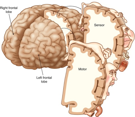
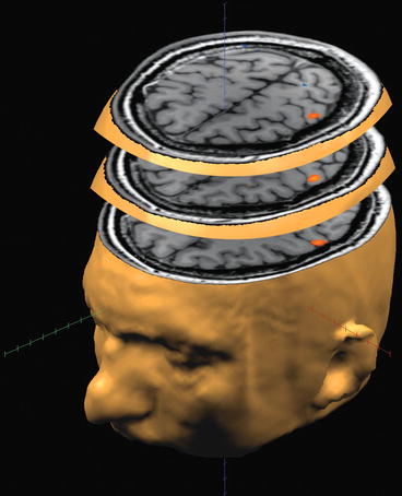
Fig. 8.3
The cortical body map as described by Penfield and Boldrey as early as 1937. The brain is seen obliquely from the frontal aspect, the arrow pointing forwards (anteriorly) and the areas for sensory and motor functions have been extracted as separate three-dimensional sections. The somatosensory cortical area is situated immediately posterior to the area for motor functions, the two cortical areas being separated by the central sulcus. The hand is represented in very large areas of the somatosensory and motor cortices. The hand representation is located close to the face. The thumb is especially big in this area and is located closest to the face
Fig. 8.4


Functional magnetic resonance imaging (fMRI) technology can be used to define the cortical representations of individual fingers in the somatosensory cortex. In this picture, the brain has been sliced in horizontal sections, and the representational areas for the right thumb (bottom slice), index finger (middle slice) and little finger (upper slice) have been activated by touching these fingers (Courtesy of Andreas Weibull, Medical Radiation Physics, Malmö, Skåne University Hospital, Malmö, Lund University)
Related posts:

Stay updated, free articles. Join our Telegram channel
Full access? Get Clinical Tree
