Trauma of the Nervous System: Peripheral Nerve Trauma

Chapter 50D Trauma of the Nervous System


Peripheral Nerve Trauma


Historical interest in peripheral nervous system (PNS) trauma dates back many centuries. Leonardo Da Vinci made detailed anatomical drawings of the brachial plexus, believing this complex of nerves existed to ensure continued function of the upper extremity should one of its elements be severed (e.g., by a sword thrust). By 1885, Duchenne, Erb, and Klumpke all had recorded their landmark descriptions of various brachial plexus injuries. The U.S. Civil War and both World Wars provided the substrate for the systematic study of peripheral nerve injuries by 19th-century American neurologist Weir Mitchell and 20th-century neurosurgeon Barnes Woodhall, respectively; their work led to the advent of nerve injury classification systems by Sir Herbert Seddon, Sir Sydney Sunderland, and George E. Omer.


Today, up to 5% of all admissions to level I trauma centers have a peripheral nerve, nerve root, or plexus injury (Noble et al., 1998). In general, injuries to the upper extremity are more common than those to the lower extremity, accounting for two-thirds of all peripheral nerve injuries. Of the four major peripheral nervous system plexuses—cervical, brachial, lumbar, and sacral—the brachial plexus is by far the most commonly affected.


This chapter begins with a review of relevant neuroanatomy. Then the common mechanisms that cause peripheral nerve injury in adults are examined, and an approach to the diagnosis and management of such injuries is presented. Next, current surgical techniques used in nerve repair are described, followed by a look at new therapeutic technologies.


Anatomy of the Spinal Nerves of the Peripheral Nervous System


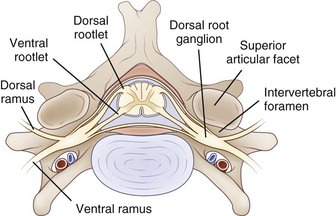
The PNS is composed of those neural elements that extend between the central nervous system (CNS—in this case the spinal cord) and their target organs (Fig. 50D.1). The peripheral motor system axons (somatic efferents) originate from the anterior horn cells and exit the spinal cord to form the ventral (anterior) rootlets. Axons of the peripheral sensory system (somatic afferent) extend from specialized sensory organs within skin, muscle, and viscera to their cell bodies, the dorsal root ganglia (DRG), which lie within the bony intervertebral foramen. These sensory fibers make up the dorsal (posterior, somatic afferent) rootlets that enter the posterior horn of the spinal cord. The mixed spinal nerves are formed when anterior and posterior rootlets combine within the neural foramen just distal to the DRG. The short spinal nerve then divides into two branches: (1) a large anterior branch (ventral ramus) that extends forward to supply the trunk muscles and gives rise to the roots of the plexus and (2) a small posterior branch (dorsal ramus) that extends backward to supply paravertebral muscles and skin of the neck.


Axon


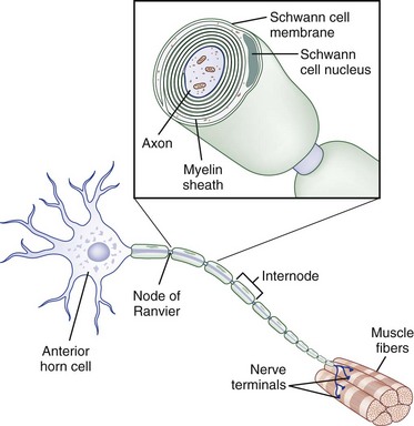
The core element of the nerve is the axon, a thin tube of axoplasm that extends from the nerve cell body to the target organ. Unmyelinated axons are partially ensheathed by invaginations of the Schwann cell membrane, whereas myelinated axons are enveloped in concentric lamellae of myelin composed of compacted spiraled Schwann cell membrane to form a sheath (Fig. 50D.2). The myelin sheath is laid down in segments called internodes, each derived from one Schwann cell. The small gap of uncovered axoplasm between sheaths is called the node of Ranvier, the site where the largest part of ion flow takes place to transmit the action potential. In saltatory conduction, action potentials leap from node to node, rather than traveling in a continuous conduction process along the entire length of the axolemma. In this way, a myelinated large-caliber axon in human adults may conduct electrical impulses at up to 73 meters per second, whereas a small unmyelinated axon may conduct as slowly as 0.5 meter per second (Kimura, 2005).


Peripheral Nerve Trunks


The connective tissue within a peripheral nerve trunk is composed of the endoneurium, perineurium, and epineurium (Fig. 50D.3). These tissues provide structure, tensile strength, and elasticity.
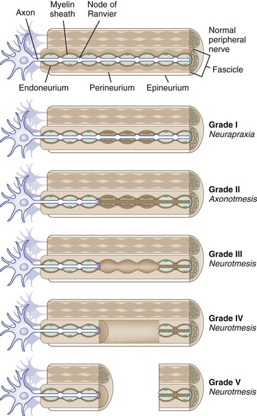


The endoneurium is a thin layer of collagenous connective tissue that surrounds individual nerve fibers and is continuous with the fine layer of connective tissue of the nerve roots. In the nerve trunk, bundles of myelinated and unmyelinated nerve fibers are arranged into fascicles (or funiculi). These fascicles, which vary greatly in size and number, are arranged in an intertwining pattern (sometimes called the Sunderland plexus) in the more proximal portion of the nerve trunk but are arrayed in a more parallel pattern in the distal parts of the nerve. Each fascicle is surrounded by perineurium, which consists of perineurial cells that interlock to form tight cell junctions, creating the blood-nerve barrier that maintains an immunologically privileged endoneurial environment. A typical spinal nerve trunk consists of a variable number of fascicles separated by inter- and extrafascicular epineurium that constitutes an extension of the dura mater. Finally, the nerve trunk melds into surrounding structures via a loose layer of protective areolar tissue, or the mesoneurium, which allows the nerve passive movement in the transverse and longitudinal planes.


Peripheral nerves have an abundant and anastomotic blood supply. The arterial arrangement is composed of extraneural arteries (arteriae nervorum) that enter the nerve proper at various sites and angles along the length of the nerve. Longitudinally arranged arterioles travel along the interfascicular epineurium before they branch off to pierce the perineurium. The endoneurial or intraneural capillaries are larger in diameter than typical capillaries in other organs and resemble end-arterioles in size, although they are surrounded only by pericytes. Venous drainage is conducted along postcapillary venules.


Classification of Nerve Trauma


Based on observations made in Great Britain in the Second World War, Seddon devised a three-tiered classification system for nerve trauma (Table 50D.1 and Fig. 50D.3). In this system, the mildest form of injury is due to a transient focal block in conduction along the nerve fiber; injury is confined to the myelin sheath and spares the axon. He called this neurapraxia (Greek for “nonaction of nerve”). This type of injury has an excellent prognosis for complete and spontaneous recovery within a 6-week period. Indeed, many patients return to normal within hours. A clinical example of neurapraxia is wrist drop secondary to prolonged external pressure that compresses the radial nerve at the spiral groove of the humerus.


Seddon’s second grade of injury, axonotmesis (Greek for “cutting of the axon”), involves injury not only to the myelin sheath but also to the axon itself so that, as noted by Seddon in 1942, “the sheath and the more intimate supporting structures of the nerve have not been completely divided, which means that the nerve as a mass of tissue is still in continuity.” Axonotmesis is common in crush injuries and displaced bone fractures, and complete recovery is less certain than with neurapraxia. It triggers the process of wallerian degeneration and regeneration, the success of which depends in part on the preservation of connective tissues such as endoneurium and perineurium.


Seddon’s most severe type of injury, called neurotmesis (a “cutting of the nerve”), entails damage to myelin, axons, and various layers of connective tissue. Seddon originally used this term to describe complete nerve transection in which “the injury produces a lesion which is in every sense complete” but later amended the definition to “describe the state of a nerve that has either been completely severed or is so seriously disorganized by scar tissue that spontaneous regeneration is out of the question.” This is common after laceration or ischemic injuries and portends the worst prognosis for clinical recovery, often necessitating surgical intervention.


A second classification system for nerve trauma was devised by Sunderland to include additional information regarding the degree of injury to connective tissue. This system is divided into five grades: grades I and II are identical to Seddon’s neurapraxia and axonotmesis, respectively (see Table 50D.1 and Fig. 50D.3). However, Sunderland subdivided Seddon’s neurotmesis into three further levels of injury: grade III entails injury to the myelin sheath, axon, and endoneurium, with sparing of the perineurium and epineurium; grade IV describes an injury to all nerve trunk elements except the epineurium; and grade V entails complete transection of all neural and connective tissue elements of the nerve trunk. Only Sunderland’s classification system is used in the rest of this chapter.


Modifications to these two systems have been devised, largely with the surgeon in mind. A further grade (grade VI) has been proposed to highlight the fact that some injuries may feature a combination of Sunderland grades (e.g., some areas may be grade III and others grade IV) affecting different fascicles within a segment of nerve.


Peripheral Nerve Degeneration and Regeneration


Large myelinated peripheral axons may respond to injury or disease in three ways: segmental demyelination, wallerian degeneration, and axonal degeneration. Segmental demyelination and wallerian degeneration are relevant to traumatic nerve injury and are discussed in more detail in this chapter, whereas axonal degeneration is more characteristically seen in metabolic and toxic nerve disorders such as diabetes mellitus and renal failure (see Chapter 76).


Segmental Demyelination


Segmental demyelination occurs when a focal segment of nerve is subjected to a relatively mild compressive or traction force. The nerve segments that lie distal and proximal to the site of injury are not affected and function normally. Conduction across the injured segment is impaired, however, when distortion of the myelin sheath causes degeneration of one or several internodes thereby reducing the ability of the sheath to act as an electrical insulator. If the myelin is only slightly damaged, the only local consequence may be a widening of the node of Ranvier that causes a slowing of conduction velocity across the nerve segment. Focal demyelination of axons within a nerve fascicle may affect some but not all fibers, resulting in asynchronous conduction across the affected nerve segment. In this case, impulses eventually reach their destination after a delay measured in milliseconds, but the slowing may affect certain nerve functions that rely on highly synchronous firing (e.g., deep tendon reflexes, vibration sense), resulting in paresthesia.


More severe compression may involve most or all myelinated nerve fibers at the injury site and several internodes. In this situation, blockade of conduction across that segment results in weakness or sensory disturbance. Study of nerve segments that have been subjected to tourniquet compression reveals that the nodes of Ranvier located at the edge of the tourniquet are subjected to greater pressure and are deformed to a greater degree than the segment lying directly beneath the tourniquet. This pressure gradient causes the underlying axon and myelin to telescope into neighboring segments, which greatly distorts the paranodal segment of myelin, resulting in conduction slowing or demyelinating conduction block.


Wallerian Degeneration


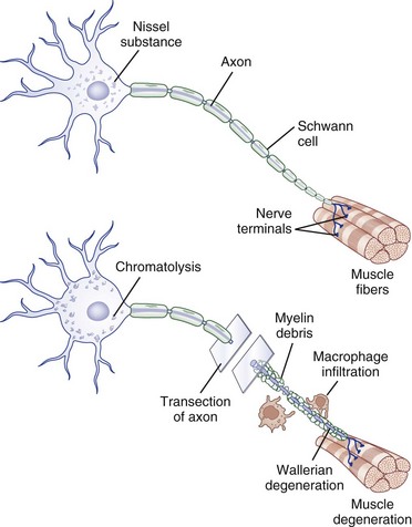
Injury to a peripheral nerve triggers a complex process that involves the axon, its cell body, its cellular connections, and the surrounding connective tissues. Wallerian degeneration follows grade II to grade V injuries and can be divided into changes that involve the segments of nerve distal and proximal to the zone of injury (Fig. 50D.4).


Distal Segment Changes


After nerve injury, the earliest changes occur in the axon distal to the site of injury, where there is disruption of retrograde and anterograde flow of signals within the axon (Makwana and Raivich, 2005). Rapid inflow of extracellular ions such as calcium and sodium occurs through the disruption in the axonal plasma membrane, which activates a cascade of events that shares features with programmed cell death or apoptosis. Axonal injury also leads to recruitment of leukocytes and initiates the cytokine-mediated signaling cascade and changes in the neighboring non-neuronal cells. This in turn triggers the synthesis of neurotrophins, chemokines, extracellular matrix molecules, proteolytic enzymes, and interleukins (Hall, 2005; Radtke and Vogt, 2009). The entire axonal process of wallerian degeneration takes approximately 1 week. By day 3, the Schwann cells retract from the node of Ranvier, and activated Schwann cells and macrophages begin to digest myelin. The process of neuronal degeneration quickly undergoes a transition to neuronal regeneration from the proximal stump. Schwann cell proliferation facilitates sprouting of new nerve branches from the injured axon terminus and initiates the sequence of events that lead to rebuilding of neural contacts to distal muscles.


Nerve Regeneration


The method of nerve regeneration depends on the type of injury sustained: remyelination after grade I lesions and collateral axon sprouting and proximal-to-distal nerve regeneration after grade II to grade V lesions.


With focal demyelinating lesions, recovery of clinical and electrodiagnostic function occurs as the Schwann cell divides and initiates remyelination. Conduction, and thereby strength, are reestablished within a few weeks or months, but the new myelin sheath usually is thinner and has several internodes for each original internode.


When only some of the axons supplying a muscle are damaged, the intact motor axons produce sprouts that reinnervate the denervated muscle fiber; this is referred to as collateral sprouting. These nerve sprouts originate from the nodes of Ranvier (nodal sprouts) or the nerve terminals (terminal sprouts) as early as 4 days from the time of injury. By adopting denervated muscle fibers, collateral innervation increases the size of the remaining motor units and results in increased contractile force. Clinical recovery due to collateral sprouting takes up to 3 to 6 months from the time of injury. During this same period, compensatory hypertrophy of muscle fibers that have retained their axons occurs, although the muscle as a whole atrophies. Enhanced synchronization of motor unit firing contributes to improved strength.


In contrast with partial or mild nerve injury, in which the surviving motor axon begins the process of collateral sprouting almost immediately, in severe or complete injury, neuronal regeneration starts from the proximal stump only after wallerian degeneration is completed (Burnett and Zager, 2004). Schwann cells play a key role in neuronal regeneration. They dedifferentiate and up-regulate the expression of adhesion molecules and neurotrophins (i.e., cadherins, immunoglobulin superfamily factors, laminin) which promote the migration of nerve sprouts that form at the regenerating axon tip. These sprouts then form cords aligned around the original basal lamina tubes of the myelinated axons (bands of Büngner) that provide a pathway along which new axons are destined to grow. The contribution of axon regeneration to clinical recovery overlaps with collateral sprouting at approximately 3 months, but axon regeneration is the main recovery mechanism from 6 to 24 months after the injury, depending in part on the distance the nerve must grow to reach its target muscle.


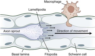
The tip of the axon sprout, called the growth cone, travels by way of filopodia and lamellipodia (Fig. 50D.5). Neurotropism, the term used to describe guidance of a regenerating axon, is accomplished by guidance molecules (i.e., semaphorins, ephrins, netrins, slits) that act to attract or repulse the growth cone to prevent misdirected growth of axon sprouts (Marx, 1995). To pass through endoneurial tubules plugged with cellular debris, the growth cone secretes plasminogen activators that dissolve cell-cell and cell-matrix adhesions. Axonal sprouts grow from the proximal to the distal stump at a rate of approximately 1 to 2 mm/day (or approximately 1 inch/month). This rate of regrowth varies depending on the location of the lesion; with proximal lesions, growth may be as fast as 2 to 3 mm/day, while that with distal lesions is about 1 mm/day.


Peripheral axons and non-neuronal cells contain growth-inducing and trophic molecules that provide the fertile milieu required for nerve regeneration. These include the neurotrophins (nerve growth factor, brain-derived neurotrophic factor, neurotrophins 3 and 4), glial cell line–derived neurotrophic factors (neurturin, artemin, persephin), insulin-like neurotrophic factor, interleukin-6, leukemia inhibitory factor, ciliary neurotrophic factor, fibroblast growth factors, and several transcription factor activators (Dieu et al., 2005; Tucker and Mearow, 2008).


The condition of the endoneurial tube at the injury site determines the efficiency of nerve regeneration. If the tube is intact, regrowth of the proximal axon has a greater chance of success. With severe or complete disruption of the endoneurial tube, the proximal axon may stray into the surrounding connective tissue, which increases the chance of disorganized nerve sprouts or neuroma formation. Misplaced axons produce ectopic hyperexcitability that results from the accumulation of sodium channels and generates neuropathic pain. Once the advancing axon has crossed the injury gap and entered the distal endoneurial tube, the nerve has a reasonably good chance of successfully reaching its target end-organ. If it takes the axon longer than 4 months to reach the gap, however, the distal endoneurial diameter shrinks to perhaps 3 µm or less, further impeding complete recovery. Nevertheless, some degree of reinnervation can occur for up to 2 years after injury.


Successful axon regeneration also relies on the presence of a viable end-organ. Denervated muscle fibers quickly undergo atrophy, and collagen deposition occurs within the endomysium and perimysium, but the overall muscle architecture is maintained, along with end-plate integrity, for at least 1 year after nerve injury. Irreversible muscle fibrosis and degeneration, however, often has occurred by 2 years. Sensory cross-reinnervation is common, and pacinian and meissnerian corpuscles (mediating rapidly adapting light touch) and Merkel cells (mediating slow-adapting touch and pressure) are able to survive in a denervated state for a period of years. Thus, even late reinnervation on the order of 2 to 3 years may restore useful protective sensory function to a limb.


Mechanisms of Traumatic Nerve injury


Compression


Compressive nerve injuries most commonly affect large-caliber myelinated nerve fibers in nerves that cross over bony surfaces or between rigid structures, such as the ulnar nerve in the ulnar groove at the elbow and the radial nerve at the spiral groove. Compression neuropathies may occur in patients who have been sedated or unconscious in a single position for an extended period. Because the primary underlying pathophysiology is that of demyelinating conduction block, an excellent recovery generally can be expected in the following days to weeks. A similar problem can be recognized in the postoperative setting. In classic postoperative brachial plexopathy, the patient awakens from general anesthesia with complete upper limb paralysis due to compression and stretch of brachial plexus elements between the clavicle and first rib while the arm is extended and hyperabducted intraoperatively. Electrophysiological examination may reveal a combination of upper trunk demyelinating conduction block and a lesser component of axon loss. The deficit characteristically reduces to a pure upper trunk distribution within the first few days, and within 2 to 3 months has fully resolved.


Mechanical compression may lead to secondary ischemic injury, as in the case of acute compartment syndromes that compromise nerve microcirculation while sparing distal-extremity pulses. When the intracompartmental pressure exceeds intraarterial pressure, nerve (and muscle) ischemia results in axon-loss lesions of variable severity. An example is the medial brachial fascial compartment syndrome after axillary arteriography or axillary regional block. Severe or irreversible single or multiple axon-loss mononeuropathies can occur within 4 hours if untreated by immediate exploration of the puncture site and decompression (Tsao and Wilbourn, 2003).


Stretch and Traction


Peripheral nerves are vulnerable to serious injury from excessive stretch or traction. As the stretching force increases, the elastic properties of the nerve are overcome, and myelin sheaths, axons, and connective tissue may rupture. In addition, it has been shown in an in vivo rat sciatic model that an 8% elongation of a nerve increases intraneural pressure to the point that blood flow is reduced by half.


An important example of a closed traction or stretch injury is neonatal Erb-Duchenne palsy due to excessive traction forces applied to the infant’s C5-C6 roots and fibers of the upper trunk of the brachial plexus during a difficult delivery. Significant axonal injury characteristically occurs, which may leave the child with permanent paralysis of upper arm flexors and forearm supinators, causing the arm to be held in the “waiter’s tip” position.


Sufficient stretch produced by distraction of the neck from the shoulder results in avulsion injury of the intraspinal rootlets from the spinal cord. Avulsion injuries most often are due to motor vehicle and motorcycle accidents. The typical scenario is that of a motorcycle rider being thrown off the motorcycle; the head and shoulder strike the pavement, laterally flexing the neck away from the shoulder, stretching the brachial plexus, and pulling the rootlets out of the spinal cord. Another example is a heavy object falling from a height onto a person’s shoulder. Avulsion particularly affects the intra- and extraforaminal portions of the cervical root fibers and the proximal brachial plexus fibers. Lesions of the lumbosacral plexus and roots are uncommon. The prognosis with avulsion of motor and preganglionic sensory fibers is poor. Avulsion is associated with particularly severe and lasting neuropathic pain.


< div class='tao-gold-member'>

Stay updated, free articles. Join our Telegram channel

Jun 19, 2016 | Posted by in NEUROLOGY | Comments Off on Trauma of the Nervous System: Peripheral Nerve Trauma

Full access? Get Clinical Tree

Get Clinical Tree app for offline access