Figure 20-1 Cross-sectional ultrasound image taken at the fibular head. The fibular head (white arrow). Hypoechogenic cyst (blue arrow). The peroneal nerve is not readily identified in this image.

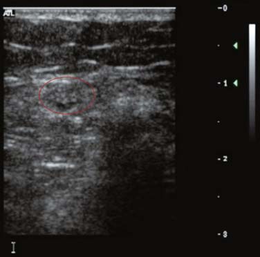
Figure 20-2 As the cyst disappears, the peroneal nerve becomes visible (outlined) and is identified by its stippled appearance.
MRI is said to be more precise than US for diagnosis of cysts at the fibular head, whereas US is typically known as a screening tool.1,2 In this case, US, performed in the electromyography laboratory as a complement to electrodiagnostic studies, played a major role in arriving at an accurate diagnosis and subsequent effective treatment of a small cyst compressing the peroneal nerve at the fibular head. This finding is not unique; US was first used to aid in diagnosis of an intraneural ganglion cyst of the peroneal nerve in 1992,3 and a recent US study found compressive intraneural peroneal ganglia in 18% of patients with peroneal mononeuropathy.4
Stay updated, free articles. Join our Telegram channel

Full access? Get Clinical Tree


