Vein of Galen Aneurysmal Malformation

CHAPTER 208 Vein of Galen Aneurysmal Malformation


Vein of Galen aneurysmal malformations (VGAMs) are arteriovenous (AV) fistulas in the choroid fissure supplied by the choroidal arteries and draining to the dilated median vein of the prosencephalon, which is the precursor of the vein of Galen and the embryonic drainage of the choroid plexus. “VGAMs” were reported to represent less than l% of all arteriovenous malformations (AVMs) in the cooperative study of subarachnoid hemorrhage.13 However, its true incidence is difficult to determine because significant diagnostic confusion exists among the various malformations that cause dilation of the vein of Galen or its embryonic precursor. The embryonic nature of the draining veins of VGAMs was first described by Raybaud and colleagues in 1989.4 The concept of this disease was further refined by Lasjaunias and associates, who subclassified VGAMs into choroidal and mural types.5,6 Other vascular lesions that cause dilation of the true vein of Galen are designated as vein of Galen aneurysmal dilation (VGAD) or vein of Galen varix (VGV). VGAD is a group of malformations associated with dilation of the vein of Galen secondary to pial or dural AV shunts draining into the true vein of Galen or its tributary. VGV is a dilated vein of Galen without AV shunts.


Classification


Choroidal Vein of Galen Aneurysmal Malformations


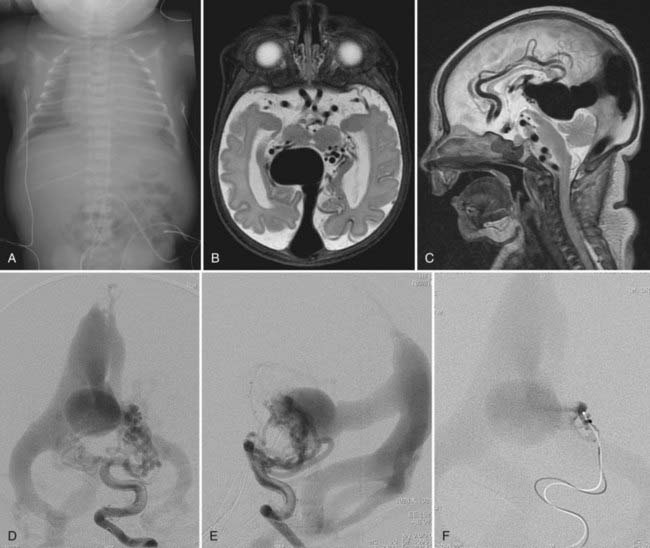
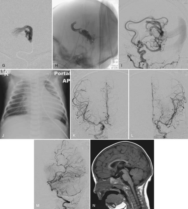
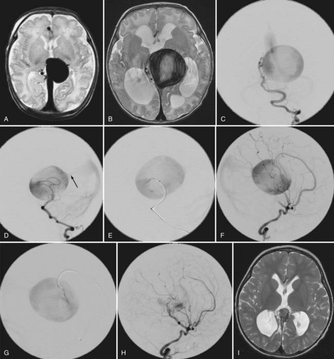
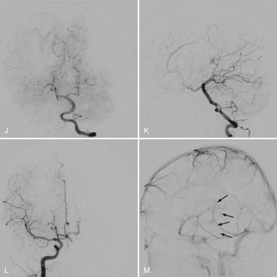
This more primitive type of VGAM consists of multiple fistulas located in the choroid fissure (Figs. 208-1 and 208-2). These multiple fistulas communicate with the anterior aspect of the median vein of the prosencephalon via an arterial network. The arterial feeders are all the choroidal arteries, including the bilateral anterior and posterior choroidal arteries, and the anterior cerebral arteries. There is often additional supply from the quadrigeminal and thalamoperforating arteries.4,7 The choroidal type of VGAM is the most severe expression of the disease and usually causes high-output cardiac failure in the newborn period because of multiple high-flow fistulas with less restriction of outflow than seen with the other type of VGAM. Choroidal VGAM is more challenging to treat because of comorbid conditions such as severe cardiopulmonary failure and the small size of the patient.


image image

FIGURE 208-1 A male infant had respiratory distress at birth. A chest radiograph at birth (A) showed marked cardiomegaly and hepatomegaly. T2-weighted axial (B) and sagittal (C) magnetic resonance imaging (MRI) at day 0 showed arteriovenous fistulas to the dilated median vein of the prosencephalon draining to the embryonic falcine sinus. Progressive congestive heart failure (CHF) developed and the infant was transferred to us on day 21. Endovascular treatment was performed on day 22. On admission, the patient was intubated with 50% FIO2 and a dobutamine drip started, as well as digoxin and furosemide (Lasix). Left vertebral artery angiograms in the posteroanterior (PA) (D) and lateral (E) views demonstrate a choroidal-type vein of Galen aneurysmal malformation (VGAM) draining to the dilated median vein of the prosencephalon and then to the embryonic falcine sinus. Multiple fistulas to the anterior portion of the median vein of the prosencephalon are supplied by bilateral posterior choroidal and thalamoperforating arteries. No straight sinus is seen. After the left vertebral angiogram, the left posterior choroidal feeder was superselectively catheterized. F, A superselective angiogram in the PA view shows multiple high-flow fistulas. They were embolized with N-butyl-2-cyanoacrylate (NBCA) under systemic hypotension.G, The cast of NBCA in the PA view. A left internal carotid angiogram after the first embolization showed the remaining fistulas supplied by the posterior choroidal arteries, the anterior choroidal artery, and the left anterior pericallosal artery (not shown). The left pericallosal artery feeders were then embolized twice with the same technique. H, The NBCA cast after the third NBCA deposition. I, A postembolization left common carotid angiogram demonstrates decreased but remaining fistulas. After this embolization procedure, the infant’s CHF improved significantly. Chest radiography 6 days after the treatment (J) showed significant improvement in his cardiomegaly. When he came back for the second procedure 6 months later, he was developing normally with normal heart and head circumference. Right (K) and left (L) common carotid angiogram in the PA view and a left vertebral angiogram in the lateral view (M) showed complete occlusion of the VGAM. T1-weighted sagittal MRI 6 months after treatment (N) showed thrombosis and shrinkage of the median vein of the prosencephalon.


image image image

FIGURE 208-2 Ten-day-old full-term baby boy in whom significant heart failure developed shortly after birth. Computed tomography and magnetic resonance imaging of the brain demonstrated a vein of Galen aneurysmal malformation (VGAM). He also had an atrial septal defect (ASD) and anomalous venous return to the heart. Emergency endovascular treatment was performed because his heart failure continued to deteriorate. At the time of treatment, he was taking furosemide (Lasix) and digoxin with worsening metabolic acidosis and oliguria. Left vertebral angiograms in the posteroanterior (PA) (A) and lateral (B) views show a choroidal-type VGAM supplied by bilateral posterior choroidal arteries and drained by the embryonic falcine sinus. There is small indirect supply from the posterior thalamoperforating arteries (arrows in A). Right internal carotid angiograms in the PA (C) and lateral (D) views show the VGAM to be supplied by the right anterior and posterior choroidal arteries and the anterior and posterior pericallosal arteries. Left internal carotid angiograms in the PA (E) and lateral (F) views show the VGAM to be supplied by the left anterior and posterior choroidal arteries and the anterior and posterior pericallosal arteries. He underwent two embolization procedures for the VGAM in the newborn period. He also underwent surgical correction of the ASD and the anomalous venous return at 5 months of age. He was brought for the third embolization for the VGAM at 6 months of age. At this point, he had failure to thrive with tachycardia and tachypnea. He had no focal motor weakness but required digoxin, Lasix, and positive airway pressure. Right vertebral artery angiograms before the third embolization in the PA (G) and lateral (H) views show a significant increase in supply from the posterior thalamoperforating arteries (compare with A and B).Right internal carotid angiograms in the PA (I) and lateral (J) views show a significant increase in supply from the anterior thalamoperforating arteries and distal internal carotid perforators (compare with C and D). Left common carotid angiograms in the PA (K) and lateral (L) views show decreased size of the feeders from the anterior and posterior cerebral arteries but increased size of the anterior thalamoperforating arteries (compare with E and F). He underwent further embolization. The glue cast after the third embolization is shown in the PA (M) and lateral (N) views. After the third embolization, his heart failure improved significantly. When he came back for the fourth embolization 13 months after the third embolization, he was developing normally without heart failure and no medication. Left vertebral angiograms before the fourth embolization in the PA (O) and lateral (P) views show a significant decrease in the size of the median vein of the prosencephalon with minimal remaining fistula from the right posterior cerebral artery feeders. The supply from the posterior thalamoperforating arteries had regressed as a result of previous embolization of the other feeders. Q, A right common carotid angiogram in the PA view shows minimal remaining fistulas from the right posterior cerebral artery feeders. No remaining supply was seen from the right anterior circulation. R, A left common carotid angiogram in the PA view shows no supply from the left anterior circulation. The patient underwent the fourth embolization with almost complete occlusion of the fistula. Left vertebral angiograms in the PA (S) and lateral (T) views 20 months after the last embolization show complete occlusion of the fistula. The patient remained neurologically normal.


Mural Vein of Galen Aneurysmal Malformations


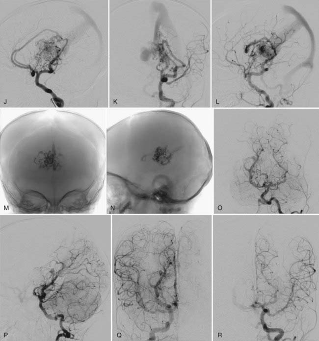
In mural VGAM, the fistulas are single or multiple and located at the wall of the dilated median vein of the prosencephalon, usually at its inferolateral margin (Fig. 208-3). The vessels supplying the shunt are usually the quadrigeminal or the posterior choroidal arteries (or both) and may be unilateral or bilateral. In contrast to choroidal-type VGAM, they have fewer fistulas and typically have more restriction of outflow, which causes more dilation of the median vein of the prosencephalon but protects the heart from high-output failure. The mural type of VGAM is therefore initially manifested later in infancy as macrocephaly, hydrocephalus, or failure to thrive, although it may be associated with mild cardiac failure or asymptomatic cardiomegaly.


image image

FIGURE 208-3 This 2-month-old boy experienced respiratory distress immediately after birth and was treated with oxygen. T2-weighted axial magnetic resonance imaging (MRI) in the newborn period (A) showed a dilated central vein and the embryonic falcine sinus without hydrocephalus. Congestive heart failure did not develop but progressive hydrocephalus did. A decision was made to perform embolization at 2 months of age. B, T2-weighted axial MRI at 2 months of age before embolization shows significant enlargement of the central vein and ventriculomegaly. Left vertebral artery angiograms in the posteroanterior (PA) (C) and lateral (D) views show a mural-type vein of Galen aneurysmal malformation (VGAM) supplied by the right lateral posterior choroidal artery. There is stenosis with restriction of venous outflow in the proximal embryonic falcine sinus (arrow in D). E, Superselective catheterization of this feeder in the lateral view, which was embolized with N-butyl-2-cyanoacrylate (NBCA) glue. F, A right internal carotid angiogram in the lateral view shows a remaining fistula supplied by the anterior pericallosal artery. G, Superselective catheterization of this feeder in the lateral view, which was also embolized with NBCA glue. H, Right common carotid lateral angiogram after the second embolization showing a small shunt remaining. I, T2-weighted axial MRI 4 years later showing thrombosis and shrinkage of the median vein of the prosencephalon. Ventricular size also decreased without ventriculoperitoneal shunting.Vertebral PA (J) and lateral (K) angiograms 4 years after treatment show complete occlusion of the VGAM. L, A right common carotid angiogram in the PA view also shows complete occlusion of the VGAM. M, The venous phase of the right common carotid angiogram in the lateral view shows no opacification of the median vein of the prosencephalon or the falcine sinus. The deep structure is draining through the epsilon-shaped collateral vein (arrows). The patient is developing normally without neurological deficits.


Vein of Galen Aneurysmal Dilation


In contrast to VGAM, the midline ectatic vein in this group of lesions is the true vein of Galen, and it therefore receives drainage from the normal brain, as well as the malformation. Two types of AVMs, pial and dural, can cause VGAD.


Pial Arteriovenous Malformation with Vein of Galen Aneurysmal Dilation


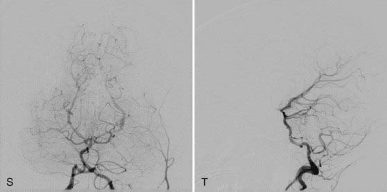
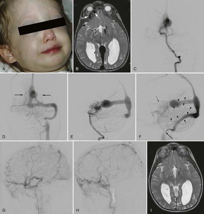
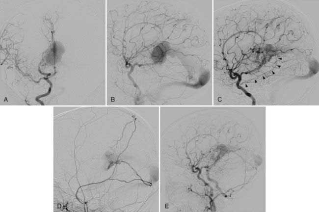
This type of VGAD is a pial or parenchymal AVM that drains into the dilated vein of Galen or its tributary (Figs. 208-4 and 208-5). Dilation of the vein of Galen is secondary to obstruction of outflow. The outflow obstruction can be relative and due to high-flow fistulas or absolute and due to progressive occlusion of the jugular bulb and the sigmoid sinus. Progressive occlusion of the dural sinus is frequently observed with pediatric fistulous malformations of the brain, including VGAM. The etiology of this outflow obstruction is unknown but may be related to underdevelopment of the jugular bulb, abnormal skull base maturation, and high-flow angiopathy of the venous system causing kinking or thrombosis at the tentorial or dural edge of the skull base. Because of this outflow restriction, the vein of Galen dilates and blood flow refluxes into other normal cerebral veins (the internal cerebral, vermian, hippocampal, basal, medial ventricular, parietal, and occipital veins or other normal tributaries of the vein of Galen). Patent embryonic sinuses such as the falcine sinus and the occipital sinus are often seen in both this type of VGAD and VGAM.


image image

FIGURE 208-4 Two-year-old girl with a tectal arteriovenous malformation and dilation of the vein of Galen. She had prominent facial veins and macrocephaly at birth and was initially managed conservatively, but seizures developed at 2 years of age, and she was referred to us for treatment. A, Clinical picture at 2 years of age showing the dilated facial veins. B, T2-weighted axial magnetic resonance imaging (MRI) showing a vascular malformation draining to a dilated central vein and the embryonic falcine sinus. There is also dilation of the right superior ophthalmic vein (arrows). The posterior horns of the lateral ventricles are dilated. Left vertebral angiograms in the posteroanterior (PA) (C and D) and lateral (E and F) views show a tectal arteriovenous malformation supplied by the posterior thalamoperforating and quadrigeminal arteries. The venous drainage is through the dilated vein of Galen and then to the embryonic falcine sinus. Reflux of venous drainage to the bilateral basal veins (arrows in D) and the striate vein (small arrow in F) proves that this is not a vein of Galen aneurysmal malformation. Late phases (D and F) show occlusion of the right sigmoid sinus and stenosis at the left jugular bulb (large arrow in F). There is also reflux of venous drainage from the torcular to the posterior fossa veins (arrowheads in F) and the superior sagittal sinus, which refluxes to the supratentorial cortical vein. Right (G) and left (H) lateral internal carotid angiograms in the late phase show a major portion of the venous drainage of both hemispheres to the superior ophthalmic veins through the cavernous sinus. She underwent three sessions of transarterial embolization with N-butyl-cyanoacrylate glue. I, T2-weighted axial MRI 10 months after the third treatment shows thrombosis and shrinkage of the vein of Galen.Left vertebral angiograms in the PA (J and K) and lateral (L and M) views on the same day as MRI (I) show complete obliteration of the malformation with significant improvement in venous drainage of the posterior fossa. There is partial recanalization of the right sigmoid sinus and recruitment of the perimedullary vein for drainage of the posterior fossa (arrows in K and M). Also note the favorable remodeling of the left jugular bulb stenosis (compare with F). Right (N) and left (O) lateral internal carotid angiograms in the late phase show significant improvement in the venous drainage of both hemispheres. P, Clinical picture on the same day as MRI (I) showing decreased prominence of the facial veins. The patient is neurologically normal without seizures.


image

FIGURE 208-5 Six-month-old girl who had increased head circumference at the age of 2 months. Magnetic resonance imaging revealed a dilated vein of Galen and ventriculomegaly. Physical examination showed normal development with dilated veins in the face and scalp. Right common carotid angiograms in the posteroanterior (A) and lateral (B) views show mainly tectal arteriovenous fistulas to the dilated vein of Galen supplied by the anterior pericallosal artery and posterior choroidal arteries. Venous drainage is thorough the true straight sinus. No dural feeders were seen. A left vertebral angiogram revealed quadrigeminal and thalamoperforator feeders (not shown). She underwent four sessions of transarterial embolization and came back for the fifth session 9 months after the first treatment. Clinically, she has been developing normally but has macrocephaly and ventriculomegaly. The size of the ventricles decreased slightly without placement of a ventriculoperitoneal shunt. C, A right internal carotid angiogram in the lateral view before the fifth embolization shows decreased size of the vein of Galen and straight sinus with remaining posterior choroidal feeders. Dural feeders developed from the artery of the free margin of the tentorium (arrowheads). D, A right external carotid angiogram before the fifth embolization shows the development of dural feeder from the middle meningeal artery. She underwent three embolizations with N-butyl-cyanoacrylate glue from the pial feeders. E, A right common carotid angiogram 4 months later shows further decreased size of the vein of Galen and the straight sinus and decreased dural supply without embolization of the dural feeders.


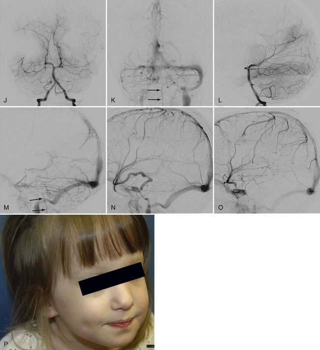
This type of VGAD is usually initially manifested in childhood or young adulthood as intracerebral hemorrhage, focal neurological deficit, or seizures. High-output cardiac failure can also occur in young children. Angiographic differentiation between VGAM and VGAD, especially a tectal AVM, can sometimes be difficult. Demonstration of transmesencephalic feeders by magnetic resonance imaging (MRI) or angiography confirms the pial nature of the lesion.8 Transmesencephalic feeders are projected below the P2 segment of the posterior cerebral artery on the lateral view of the vertebral artery angiogram.8 For treatment, transvenous occlusion of the venous dilation in the VGAD is contraindicated because it may produce hemorrhage or venous infarction of the deep cerebral structures as a result of occlusion of the outflow of these veins.


Dural Arteriovenous Malformation with Vein of Galen Aneurysmal Dilation


The dural AVM with VGAD is an acquired lesion in which AV shunts are located in the wall of the vein of Galen itself; it usually develops in the fourth or fifth decade of life. The vein of Galen dilation is due to stenosis or thrombosis of the straight sinus. Reflux is always noted into afferent cerebral veins from the vein of Galen. Typical clinical findings are headaches and progressive dementia secondary to cerebral venous hypertension. The arterial supply is derived predominantly from dural falcotentorial arteries from the internal and external carotid arteries and the vertebral artery, as well as the vasa vasorum to the wall of the vein of Galen from the pial arteries.9 Endovascular treatment of this type of dural AVM had been difficult because the transvenous approach is often not feasible and there are too many feeders from both the carotid and vertebral arteries for transarterial embolization. The recent introduction of Onyx (ev3, Irvine, CA) for embolization of dural AVMs has significantly improved the results of treatment of this type of dural AVM because a large volume of Onyx can be injected through one feeder to occlude an extensive dural vascular network supplied by multiple feeders and shunting to the dilated vein of Galen.


Embryology


The choroid plexus develops while the brain parenchyma is still not penetrated by vessels and is nurtured by diffusion from the surrounding vascularized meninx primitiva. Development of the choroid plexus is accompanied by the development of arteries and veins supplying and draining the choroid plexus. Arteries to the choroid plexus include the anterior cerebral artery, anterior choroidal artery, and posterior choroidal artery. At this developmental stage, the quadrigeminal arteries develop during growth of the quadrigeminal plate. The quadrigeminal arteries are numerous and connected by a meningeal capillary network in the meninx primitiva, similar to the pattern seen in VGAMs. Thus, as early as the fifth week, the first arteries to differentiate are the choroidal and quadrigeminal arteries, which are the primary arterial supply of the VGAM.


Expansion of the choroid plexus on the roof of the diencephalon induces the development of a midline dorsal vein draining the bilateral choroid plexuses. This vein is the first vein to drain an intracerebral structure and is designated the median vein of the prosencephalon. This vein remains functional during the second month and the first half of the third month of development. Progression of intracerebral vascularization and development of the basal ganglia result in formation of the paired internal cerebral veins, which then annex the venous drainage of the choroid plexus. This leads to regression of the median vein of the prosencephalon except for the most caudal portion, which joins with the internal cerebral veins to form the vein of Galen. Based on analysis of the vascular anatomy of VGAMs and vascular embryology, Raybaud and colleagues estimated that formation of the VGAM probably occurs between the embryonic stage of 21 to 23 mm (6 weeks) and 50 mm (11 weeks).4


Angioarchitecture of Vein of Galen Aneurysmal Malformations


VGAM, including the feeding arteries and the draining vein, exists in the subarachnoid space within the choroid fissure. The choroid fissure consists of the cistern of the velum interpositum and the quadrigeminal cistern. The arteries in the cistern of the velum interpositum are the choroidal arteries, including the anterior and posterior choroidal arteries, and the anterior cerebral arteries. The posterior choroidal arteries can have anastomoses among themselves without the existence of a VGAM. The anterior choroidal artery reaches the foramen of Monro along the choroid plexus. The anterior cerebral artery courses around the splenium of the corpus callosum anteriorly and supplies the choroid plexus at the foramen of Monro. The arteries in the quadrigeminal cistern are quadrigeminal (collicular) arteries coming off the posterior circle of Willis. They usually originate from the crural or ambient segment of the posterior cerebral artery, the superior cerebellar artery, and the medial posterior choroidal artery. They form a dense arterial network in the subarachnoid space above the quadrigeminal plate.4


Other potential contributors to the VGAM include thalamoperforating and subependymal feeders from the posterior circle of Willis. They are secondary feeders because of the “sump” effect of the venous drainage and may regress after occlusion of the primary shunts (see Fig. 208-2). Dural feeders may exist or develop secondarily via the supply to the vasa vasorum at the venodural junction. The choroidal-type VGAM may form fistulas to the choroidal vein at the interventricular foramen and is supplied by perforators from the anterior cerebral artery or Heubner’s artery.7


The dilated draining vein of the VGAM is the embryonic median vein of the prosencephalon, as described in the embryology section. It is located in the midline and receives bilateral supply, although it may be shifted to one side, opposite the major fistula. A choroidal VGAM is more primitive and has multiple fistulas with an interposing arterial network between the feeders and the draining vein. The mural type has single or multiple fistulas on the wall of the dilated draining vein. Multiple feeders can converge into a common vascular channel opening into the dilated vein. A mixed malformation with features of both the choroidal and mural types may also occur.7


The median vein of the prosencephalon has no communication with the drainage system of the normal brain. The choroidal veins embryologically drain into the median vein of the prosencephalon and may remain connected to the median vein with or without the existence of fistulas to the choroidal vein itself. This connection is seen mostly with choroidal-type VGAMs and may mediate communication between the median vein of the prosencephalon and the striate vein, which may be a cause of hemorrhage or venous infarction after transvenous embolization of the VGAM. The straight sinus is absent with most VGAMs, and the dilated median vein of the prosencephalon usually drains to the embryonic falcine sinus, which connects to the posterior third of the superior sagittal sinus.10 Persistence of other embryologic sinuses, such as the occipital and marginal sinuses, is often observed in patients with VGAMs. Persisting arterial anomalies such as a limbic arterial ring involving the anterior and posterior choroidal arteries and pericallosal arteries are also frequently present.


Because of lack of connection to the median prosencephalic vein, the deep venous drainage of the brain takes an alternative pathway. The most commonly observed pattern of deep venous drainage is through the thalamic vein connecting to the lateral mesencephalic vein. This drainage system takes an epsilon shape and is seen to connect to the superior petrosal sinus on lateral angiographic views.10,11

Only gold members can continue reading. Log In or Register to continue

Stay updated, free articles. Join our Telegram channel

Aug 7, 2016 | Posted by in NEUROSURGERY | Comments Off on Vein of Galen Aneurysmal Malformation

Full access? Get Clinical Tree

Get Clinical Tree app for offline access