Ventral/Lateral Paraspinal Mass
Jeffrey S. Ross, MD
DIFFERENTIAL DIAGNOSIS
Common
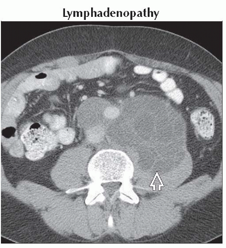
Lymphadenopathy
Lymphoma
Metastases
Metastases, Vertebral Body
Aortic Aneurysm
Paraspinal Abscess
Retroperitoneal Hemorrhage
Meningocele, Lateral
Neurogenic Tumor
Schwannoma
Neuroblastoma
Ganglioneuroma
Less Common
Extramedullary Hematopoiesis
Retroperitoneal Lymphocele
Retroperitoneal Fibrosis
Liposarcoma, Soft Tissue
Fibrosarcoma, Soft Tissue
ESSENTIAL INFORMATION
Helpful Clues for Common Diagnoses
Lymphadenopathy
Lymphatic or hematogenous spread
Testicular, ovarian cancer
Melanoma
Prostate, lung, breast
Direct extension from primary intra-abdominal neoplasms: Pancreas, GI cancers
Testicular germ cell tumors: Nodes at level of ipsilateral renal hilum
Metastases
Vertebral body involvement with sparing of disc space, with adjacent soft tissue extension
Aortic Aneurysm
Dilatation of the aorta (Ao) > 1.5x the normal diameter (5 cm ascending Ao, 4 cm arch/thoracic Ao, 3 cm distal abdominal Ao)
Always measure Ao outer-to-outer borders
Symptoms: Chest, back, or abdominal pain, distal embolization
Paraspinal Abscess
Look for associated disc space infection
Central necrotic foci without enhancement
Retroperitoneal Hemorrhage
High density collection in retroperitoneal space with fluid-fluid level
Causes include anticoagulation, aneurysm, & tumor rupture
Meningocele, Lateral
CSF signal/density meningeal protrusion through neural foramen into adjacent intercostal/extrapleural space
Strong association with NF1
Neurogenic Tumor
Schwannoma: Well-circumscribed, “dumbbel”-shaped, enhancing spinal mass centered about foramen
Image Gallery
 Axial CECT shows a metastatic germ cell tumor as a large confluent low density retroperitoneal mass
 that displaces the aorta anteriorly. that displaces the aorta anteriorly.Stay updated, free articles. Join our Telegram channel
Full access? Get Clinical Tree
 Get Clinical Tree app for offline access

|