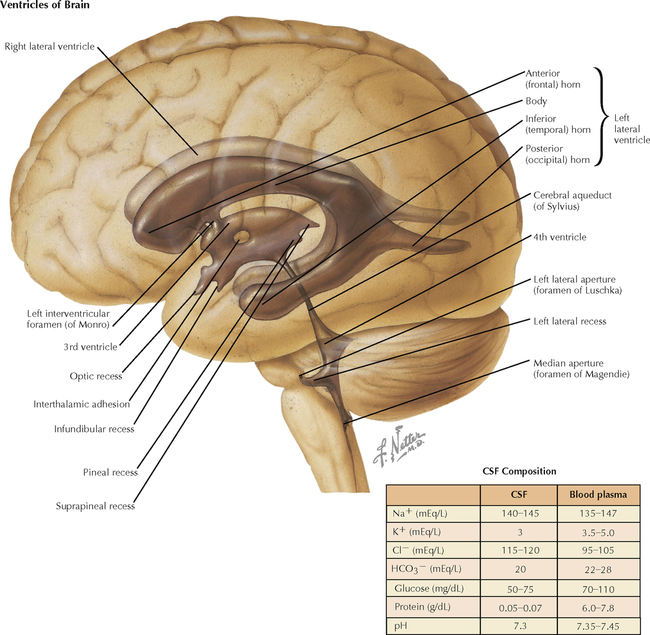
6 VENTRICLES AND THE CEREBROSPINAL FLUID 6.1. Ventricular Anatomy 6.2. Ventricular Anatomy in Coronal Forebrain Section 6.3. Anatomy of the Fourth Ventricle: Posterior View with Cerebellum Removed 6.4. Anatomy of the Fourth Ventricle: Lateral View 6.5. Magnetic Resonance Imaging of the Ventricles: Axial and Coronal Views 6.6. Circulation of the Cerebrospinal Fluid 6.1 VENTRICULAR ANATOMY The lateral ventricles are C-shaped, reflecting their association with the developing telencephalon as it sweeps upward, back, and then down and forward as the temporal lobe. The position of the lateral ventricles in relation to the head and body of the caudate nucleus is an important radiological landmark in a variety of conditions, such as hydrocephalus, caudate atrophy in Huntington’s disease, and shifting of the midline with a tumor. Cerebrospinal fluid (CSF) flows through the interventricular foramen of Monro into the narrow third ventricle, then into the cerebral aqueduct and the fourth ventricle. Blockage of flow in the aqueduct can precipitate internal hydrocephalus, with swelling of the ventricles rostral to the site of blockage. The escape sites where CSF can flow into expanded regions of the subarachnoid space called cisterns are the medial foramen of Magendie and the lateral foramina of Luschka. These foramina are additional sites where blockage of CSF flow can occur. The choroid plexus, extending into the ventricles, produces the CSF. 6.2 VENTRICULAR ANATOMY IN CORONAL FOREBRAIN SECTION A coronal section through the diencephalon shows the bodies of the lateral ventricles, the narrow interventricular foramina of Munro, and the midline third ventricle. The flow of CSF is from the lateral ventricles into the third ventricle. The choroid plexus protrudes into both the lateral and third ventricles and produces CSF. The temporal (inferior) pole of the lateral ventricle and its associated choroid plexus is shown in the temporal lobe. Only gold members can continue reading. Log In or Register to continue Share this:Click to share on Twitter (Opens in new window)Click to share on Facebook (Opens in new window) Related Related posts: SPINAL CORD MOTOR SYSTEMS NEURONS AND THEIR PROPERTIES AUTONOMIC-HYPOTHALAMIC-LIMBIC SYSTEMS TELENCEPHALON PERIPHERAL NERVOUS SYSTEM Stay updated, free articles. Join our Telegram channel Join Tags: Netters Atlas of Neuroscience with STUDENT CONSULT Online Access Jun 4, 2016 | Posted by admin in NEUROLOGY | Comments Off on VENTRICLES AND THE CEREBROSPINAL FLUID Full access? Get Clinical Tree
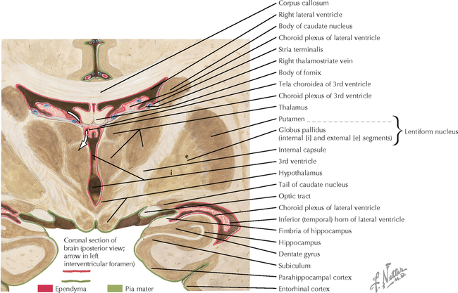
6 VENTRICLES AND THE CEREBROSPINAL FLUID 6.1. Ventricular Anatomy 6.2. Ventricular Anatomy in Coronal Forebrain Section 6.3. Anatomy of the Fourth Ventricle: Posterior View with Cerebellum Removed 6.4. Anatomy of the Fourth Ventricle: Lateral View 6.5. Magnetic Resonance Imaging of the Ventricles: Axial and Coronal Views 6.6. Circulation of the Cerebrospinal Fluid 6.1 VENTRICULAR ANATOMY The lateral ventricles are C-shaped, reflecting their association with the developing telencephalon as it sweeps upward, back, and then down and forward as the temporal lobe. The position of the lateral ventricles in relation to the head and body of the caudate nucleus is an important radiological landmark in a variety of conditions, such as hydrocephalus, caudate atrophy in Huntington’s disease, and shifting of the midline with a tumor. Cerebrospinal fluid (CSF) flows through the interventricular foramen of Monro into the narrow third ventricle, then into the cerebral aqueduct and the fourth ventricle. Blockage of flow in the aqueduct can precipitate internal hydrocephalus, with swelling of the ventricles rostral to the site of blockage. The escape sites where CSF can flow into expanded regions of the subarachnoid space called cisterns are the medial foramen of Magendie and the lateral foramina of Luschka. These foramina are additional sites where blockage of CSF flow can occur. The choroid plexus, extending into the ventricles, produces the CSF. 6.2 VENTRICULAR ANATOMY IN CORONAL FOREBRAIN SECTION A coronal section through the diencephalon shows the bodies of the lateral ventricles, the narrow interventricular foramina of Munro, and the midline third ventricle. The flow of CSF is from the lateral ventricles into the third ventricle. The choroid plexus protrudes into both the lateral and third ventricles and produces CSF. The temporal (inferior) pole of the lateral ventricle and its associated choroid plexus is shown in the temporal lobe. Only gold members can continue reading. Log In or Register to continue Share this:Click to share on Twitter (Opens in new window)Click to share on Facebook (Opens in new window) Related Related posts: SPINAL CORD MOTOR SYSTEMS NEURONS AND THEIR PROPERTIES AUTONOMIC-HYPOTHALAMIC-LIMBIC SYSTEMS TELENCEPHALON PERIPHERAL NERVOUS SYSTEM Stay updated, free articles. Join our Telegram channel Join Tags: Netters Atlas of Neuroscience with STUDENT CONSULT Online Access Jun 4, 2016 | Posted by admin in NEUROLOGY | Comments Off on VENTRICLES AND THE CEREBROSPINAL FLUID Full access? Get Clinical Tree