Vertebral Body Scalloping/Widened Canal
Bryson Borg, MD
DIFFERENTIAL DIAGNOSIS
Common
Normal Variant
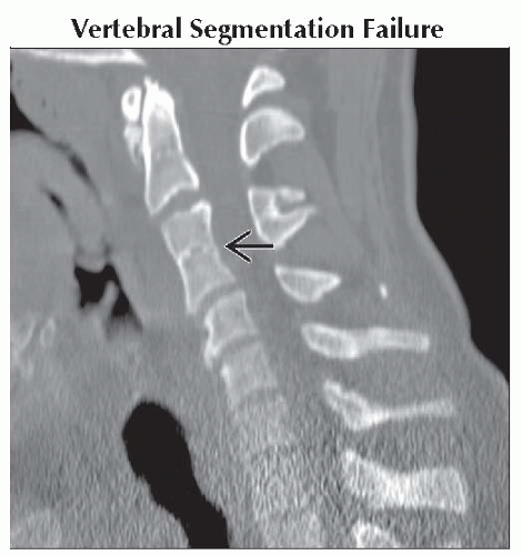
Vertebral Segmentation Failure
Dural Dysplasia
Intraspinal Mass
Ependymoma
Schwannoma
Neurofibroma
Meningioma
Arachnoid Cyst
Lipoma
Astrocytoma
Dermoid and Epidermoid Tumors
Less Common
Congenital Skeletal Disorders
Achondroplasia
Mucopolysaccharidoses (Morquio, Hurler)
Diastematomyelia
Juvenile Idiopathic Arthritis
Severe, Longstanding Communicating Hydrocephalus
Hydrosyringomyelia
Acromegaly
ESSENTIAL INFORMATION
Helpful Clues for Common Diagnoses
Normal Variant
Body slightly concave on all surfaces
Vertebral Segmentation Failure
Single level cervical fusion a rather common, incidental anomaly
“Waisted” appearance at the fusion due to stress shielding
Dural Dysplasia
Intrinsic weakness in dura, transmitted CSF pressure causes bony remodeling
Seen with neurofibromatosis type 1, Marfan disease, homocystinuria, Ehlers-Danlos, and ankylosing spondylitis
Ependymoma
Enhancing intramedullary or intradural-extramedullary (lumbosacral canal) mass
Schwannoma
Extramedullary mass, typically ventral or lateral to the cord or within fibers of the cauda equina
Those associated with canal remodeling typically fusiform or dumbbell in shape
Neurofibroma
In this context, not reliably differentiated from schwannoma (see above)
Meningioma
Intradural-extramedullary enhancing mass, with broad dural base
Arachnoid Cyst
Circumscribed, thin-walled, nonenhancing extramedullary mass, follows CSF signal/attenuation
Helpful Clues for Less Common Diagnoses
Achondroplasia
Congenitally short, narrowed pedicles; bullet-shaped vertebral bodies with posterior scalloping
Image Gallery
 Sagittal bone CT shows developmental fusion of C3 and C4 with “waisting” at the level of the fusion
 . .Stay updated, free articles. Join our Telegram channel
Full access? Get Clinical Tree
 Get Clinical Tree app for offline access

|