Vertebral Body Sclerosis, Focal
Bryson Borg, MD
DIFFERENTIAL DIAGNOSIS
Common
Bone Island
Hemangioma
Degenerative Endplate Changes
Metastases, Blastic Osseous
Insufficiency Fracture, Pedicle
Vertebroplasty/Kyphoplasty
Compression Fracture
Less Common
Chronic Osteomyelitis
Early Ankylosing Spondylitis
Osteoid Osteoma
ESSENTIAL INFORMATION
Helpful Clues for Common Diagnoses
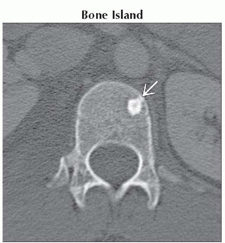
Bone Island
Most common cause of a focal sclerotic density
Cortical bone density with faint, spiculated border
Normal uptake on bone scan
Hemangioma
Coarse, thickened vertical trabeculations
Degenerative Endplate Changes
CT analog to type 3 marrow on MR
Should see advanced degenerative change in the adjacent disc space
Extensive endplate sclerosis: Consider chronic osteomyelitis
Metastases, Blastic Osseous
Multiple vertebral bodies typically involved
Sclerotic (e.g., prostate, carcinoid, medulloblastoma, neuroblastoma) or mixed sclerotic and lytic (breast, lung)
Insufficiency Fracture, Pedicle
Reactive sclerosis of portions of the neural arch associated with abnormal biomechanical loading
Can be seen with chronic fractures of the contralateral pedicle or pars, developmentally incomplete neural arch, or advanced degenerative change
Compression Fracture
Sclerosis can be seen resulting from either trabecular impaction or from the healing response
Helpful Clues for Less Common Diagnoses
Chronic Osteomyelitis
Advanced destructive endplate changes, paravertebral soft tissue &/or fluid
Typically more extensive involvement of the adjacent vertebral bodies than seen with degenerative endplate changes
Early Ankylosing Spondylitis
Erosions and reactive sclerosis result in square vertebral bodies and “shiny corners”
Osteoid Osteoma
Central lytic focus with surrounding (reactive) bony sclerosis
Classic history is night pain, relieved by aspirin/NSAIDs
Image Gallery
 Axial bone CT shows dense, cortical bone density lesion
 with characteristic appearance of a bone island, including spiculated or “brush-like” margins and absence of destructive changes. with characteristic appearance of a bone island, including spiculated or “brush-like” margins and absence of destructive changes.Stay updated, free articles. Join our Telegram channel
Full access? Get Clinical Tree
 Get Clinical Tree app for offline access

|