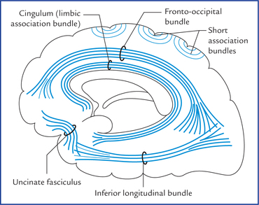
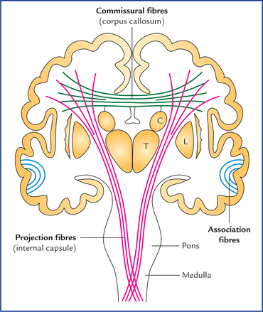
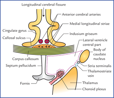
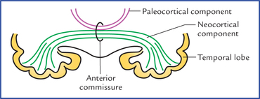
14 The cerebral white matter is a compact mass of a vast number of nerve fibres and associated neuroglia. It lies deep to the cerebral cortex and forms the large volume of each cerebral hemisphere. The fibres of white matter connect the various parts of cerebral cortex with each other and to the other parts of the central nervous system. They are classified into following three types, on the basis of the types of connections they provide (Fig. 14.1): Fig. 14.1 Frontal view of coronal section of the brain showing association, commissural, and projection fibres. (C = caudate nucleus, T = thalamus, L = lentiform nucleus.) The association fibres interconnect the different regions of the cerebral cortex in the same hemisphere (intrahemi-spheric fibres). These are of two types (Fig. 14.2): • Short association fibres (arcuate or ‘U’ fibres) which interconnect the adjacent gyri by hooking around the sulcus, hence they are also called arcuate fibres. • Long association fibres travel for long distances and interconnect the widely separated gyri, viz. gyri of different lobes. The long association fibres are grouped into bundles. The examples of bundles of long association fibres are as follows: 1. Uncinate fasciculus which connects the motor speech area and orbital cortex of frontal lobe with the cortex of temporal pole by hooking around the stem of lateral sulcus. It is narrow in the middle and fanned out at both ends. 2. Cingulum (also called limbic association bundle): It is thick bundle of fibres occupying the cingulate and parahippocampal gyri. It extends from the parater-minal gyrus to the uncus forming almost a circle like a girdle (cingulum = girdle) hence its name. 3. Superior longitudinal bundle: It is the longest association bundle which connects the frontal lobe to the occipital and temporal lobes. 4. Inferior longitudinal bundle: It connects the visual association area of occipital lobe to the temporal lobe. 5. Fronto-occipital bundle: It commences in the frontal pole, runs backwards to radiate into the occipital and temporal lobes. The fronto-occipital bundle pursues a similar course to that of superior longitudinal fasciculus. However, it lies deep to the superior longitudinal bundle and is separated from it by the fibres of the corona radiata. The important commissures of the brain are as follows: Fig. 14.3 Median sagittal section of the cerebrum showing shape and different parts of corpus callosum. External features and relations of corpus callosum • Corpus callosum forms a massive arched interhemispheric bridge in the floor of the median longitudinal cerebral fissure connecting the medial surfaces of the two cerebral hemispheres. • It forms massive, arched, interhemispheric bridge flooring the midline longitudinal fissure and roofing both the lateral ventricles. • It lies nearer the anterior end (4 cm behind the frontal pole) of the hemisphere than the posterior end (6 cm in front of the occipital pole). • In sagittal section of cerebrum it is seen as C-shaped mass of white fibres on the medial surface of the hemisphere forming the roof of the lateral ventricle (Fig. 14.4). Fig. 14.4 Some of the relations of corpus callosum in the coronal section of the brain passing through the central parts of lateral ventricles parts. Fig. 14.5 Horizontal section of the cerebrum (schematic) to show the course and components of the fibres of corpus callo-sum. Note that the fibres of corona radiata are cut at right angle to their course. • The concave inferior aspect of corpus callosum is attached with the convex superior aspect of the fornix by the septum pellucidum and its convex superior aspect is covered by a thin layer of grey matter, the indusium griseum, embedded in which are the fibre bundles of bilateral medial and lateral longitudinal striae. • The anterior cerebral vessels often lie on the pia mater covering its superior aspect of corpus callosum. • The superior aspect of corpus callosum is covered on each side by cingulate gyrus from which it is separated by a callosal sulcus. The corpus callosum is divided from before backwards into four parts: (a) rostrum, (b) genu, (c) trunk/body, and (d) splenium (Fig. 14.3). The fibres of the splenium connect the posterior parts of the parietal lobes, and temporal and occipital lobes of the two hemispheres. The fibres connecting the occipital lobes sweep backwards on either side above the calcarine sulcus forming a large fork-like structure, the forceps major (Fig. 14.5). The forceps major forms a swelling in the upper part of the medial wall of the posterior horn of lateral ventricle, the bulb of the posterior horn. Anterior commissure consists of two components (Fig. 14.6): Fig. 14.6 Anterior commissure. The large neocortical component is shown in green and the small paleocortical component in maroon. • A large posterior neocortical component, which interconnects the lower and anterior parts of the temporal lobes. • A smaller anterior paleocortical component, which interconnects the olfactory regions (olfactory bulbs, olfactory tubercles, etc.) of the two hemispheres. Seen from below the full extent of anterior commissure has the shape of a cupid’s bow. It interconnects the superior colliculi, pretectal and interstitial nuclei of two sides.
White Matter of the Cerebrum and Lateral Ventricles
White Matter Of The Cerebrum
Types of Fibres in the White Matter
Association fibres
Commissural fibres
Corpus callosum (Figs 14.3–14.5)
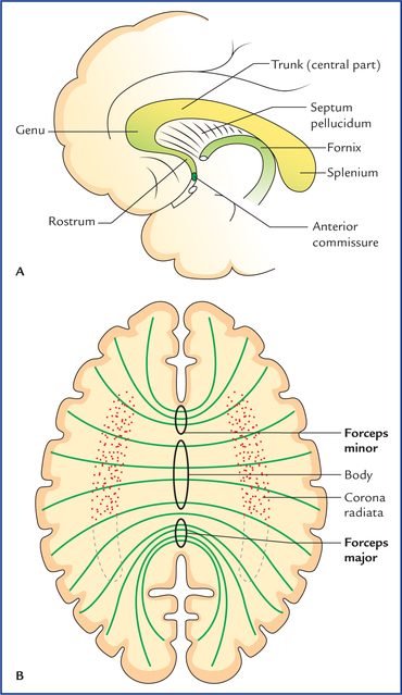
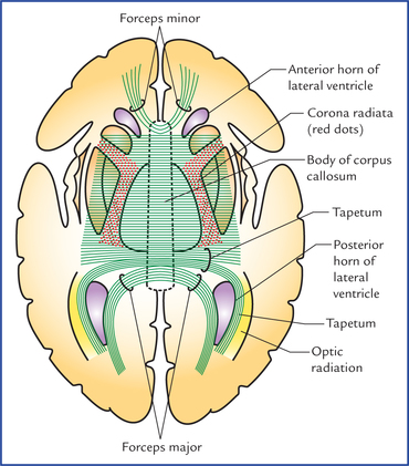
Parts of the corpus callosum
Anterior commissure
Posterior commissure
![]()
Stay updated, free articles. Join our Telegram channel

Full access? Get Clinical Tree


White matter of the cerebrum and lateral ventricles 14