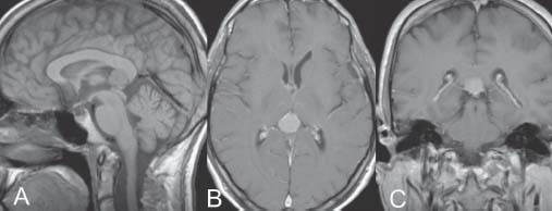
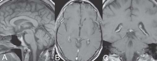
Case 10 Velum Interpositum Meningioma Fig. 10.1 (A) Sagittal, (B) axial, and (C) coronal T1-weighted magnetic resonance images with gadolinium. Fig. 10.2 Postoperative (A) sagittal, (B) axial, and (C) coronal T1-weighted magnetic resonance images with gadolinium.
 Clinical Presentation
 Questions
 Answers
< div class='tao-gold-member'>
Only gold members can continue reading. Log In or Register to continue


