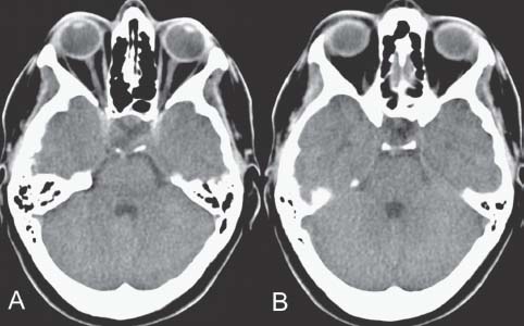
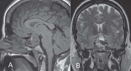
11 Pituitary Apoplexy

Case 11 Pituitary Apoplexy


Michel W. Bojanowski and Denis Klironomos


Image

Fig. 11.1 (A,B) Head computed tomography (CT) scan showing axial cuts through the sella turcica.


Image Clinical Presentation


Image Questions


  1. Interpret the CT scan.
  2. What is your initial diagnosis and why?
  3. What are important questions to ask when obtaining the history and points to look for on the physical examination?
  4. What is your initial management?

    The headaches have gradually decreased over the next few hours. A magnetic resonance imaging (MRI) scan of the brain is obtained (Fig. 11.2).


  5. Describe the MRI. What is your management now?
  6. What are the indications for surgery?

    An MRI is obtained a few weeks later (Fig. 11.3).


  7. Discuss the findings.
  8. What is the pathophysiology of this condition?
  9. What are the precipitating factors?
  10. What is the expected outcome after appropriate management?


Image

Fig. 11.2 (A) Sagittal T1-weighted and (B) coronal T2-weighted weighted magnetic resonance imaging of the brain at the level of the sella.


Image

Fig. 11.3 (A) Sagittal T1-weighted magnetic resonance imaging (MRI) scan without gadolinium and (B) coronal T1-weighted MRI scan with gadolinium at the level of the sella.


Image Answers


  1. Interpret the CT scan.

    • The CT scan reveals significant enlargement of the pituitary fossa suggestive of an intrasellar tumor. There is no evidence of subarachnoid blood.

  2. What is your initial diagnosis and describe why.

  3. What are the important questions to ask when obtain ing the history and points to look for on the physical examination?

    • The history should include questions regarding symptoms and signs resulting from sudden and fulminant expansion of the pituitary tumor (which may be due to hemorrhage, infarction, or hemorrhagic infarction).
    • Hypocortisolism resulting from destruction or compression of the pituitary gland
    • Impairment of visual acuity or fields and ophthalmoplegia resulting from compression of neural structures when the sudden enlargement is superior or lateral
    • Meningismus due to leakage of blood and necrotic tissue in the subarachnoid space
    • Look for precipitating factors (refer to Question 9).
    • Symptoms and signs related to the presence of a secreting or nonsecreting (hypopituitarism) pituitary tumor
    • Differential diagnosis involves most commonly subarachnoid hemorrhage, followed by meningitis.1,5

  4. What is your initial management?

  5. Describe the MRI. What is your management now?

    • The MRI (Fig. 11.2) reveals a pituitary tumor with a suprasellar extension containing mixed intensities suggestive of an acute intratumoral hemorrhage.
    • Neurologic and neuro-ophthalmologic examinations are normal, and a large part of the tumor appears necrotic. Hence, conservative management is justified: obtain follow-up MRI in a few weeks provided the patient remains stable.

  6. What are the indications for surgery?

    • Opinions may vary among different authors.
    • Urgent decompression is required for sudden onset of blindness, for progressive deterioration of vision, or for neurologic deterioration due to hydrocephalus.
    • Surgery is indicated for decreased level of consciousness and for hypothalamic dysfunction. It is also required for impairment of visual acuity, constriction of visual fields, or progressive deterioration of oculomotricity. For visual impairment, surgery is recommended within the first week.3
    • Ventricular drainage may be necessary in the presence of hydrocephalus.

  7. Discuss the findings.

    • The MRI (Fig. 11.3) reveals a complete disappear ance of the tumor. Sporadic cases of pituitary apoplexy cured by isolated medical treatment have been reported.6

  8. What is the pathophysiology of this condition?

    • This is controversial. For the majority of cases, it results from hemorrhage, infarction, or hemorrhagic infarction of a pituitary tumor secondary to its rapid growth.1 This may be due to a discrepancy between the rate of neoplastic progression and the availability of circulatory input. However, although pituitary apoplexy typically occurs in pituitary macroadenomas, small tumors also hemorrhage.7 One theory suggests intrinsic vasculopathy of the pituitary adenoma with secondary susceptibility to infarction and hemorrhage.8
    • Most patients had an undiagnosed pituitary adenoma at the time of apoplexy presentation.9 Although pituitary apoplexy occurs most of the time in pituitary adenomas, it may also occur in1,2

      • Healthy pituitary gland
      • Pituitary abscess
      • Metastatic tumor
      • Lymphocytic hypophysitis
      • Craniopharyngioma

  9. What are the precipitating factors?

    • Precipitating factors are identified in ~50% of cases.4 There is no preponderance of tumor type.10 Precipitating factors that have been involved include10

      • Treatment with bromocriptine
      • Treatment with anticoagulants
      • Stimulation tests of hormonal therapy with gonadotrophin-releasing hormone agonists
      • Thrombocytopenia
      • Reduced blood flow in the gland: head trauma, recent surgery
      • Pregnancy

  10. What is the expected outcome after appropriate management?

    • Pituitary apoplexy is a potentially life-threatening condition, but the overall outcome with appropriate management is good.5,9

      • Visual acuity: Outcome is related to duration, severity of the initial defect, appearance of the optic disk, and timing of decompression.1,11 However, even complete blindness may have remarkable improvement if surgical decompression is undertaken early.12
      • Ophthalmoparesis is reported to have good outcome whether treated conservatively or with surgical decompression.8
      • Endocrine function: The majority of patients require endocrine replacement therapy.1,3,8,9

    • Thickening of the sphenoid sinus mucosa during acute pituitary apoplexy may represent an indirect measure of increased intrasellar pressure. This finding has been associated with higher grades of apoplexy, larger tumors with compression of parasellar structures, and worse endocrinologic and neurologic outcomes.13
    • Recurrent apoplexy has been documented in patients managed conservatively after their first apoplectic event but rarely after surgical treatment of the initial episode.3
< div class='tao-gold-member'>

Only gold members can continue reading. Log In or Register to continue

Stay updated, free articles. Join our Telegram channel

Jul 16, 2016 | Posted by in NEUROSURGERY | Comments Off on 11 Pituitary Apoplexy

Full access? Get Clinical Tree

Get Clinical Tree app for offline access