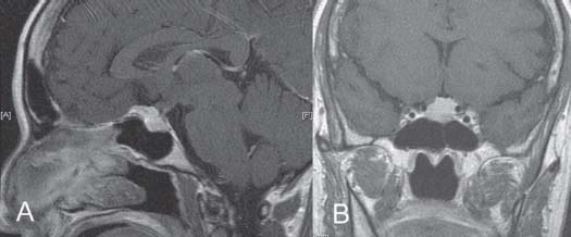
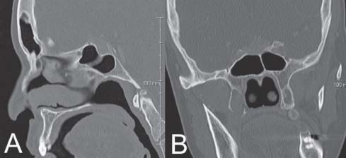
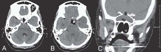
12 Pituitary Adenoma

Case 12 Pituitary Adenoma


Remi Nader and Marc-Elie Nader


Image

Fig. 12.1 (A) Sagittal and (B) coronal T1-weighted magnetic resonance images with contrast demonstrating lesion at the level of the sella.


Image Clinical Presentation


Image Questions


  1. Interpret the MRI.
  2. What other symptoms/signs do you suspect in this patient, and what other investigative measures would you like to obtain?
  3. What is your differential diagnosis?

    You obtain a computed tomography (CT) scan (Fig. 12.2) after having referred your patient to your otolaryngology colleague. The endocrinology workup reveals no abnormalities. The ophthalmologist confirms your examination findings. The patient wants to have this lesion treated.


  4. What are the indications for surgical intervention in this case?
  5. What approach would you use and why?

    You elect to proceed with a transnasal transsphenoidal approach to resect this tumor. Your otolaryngology colleague helps with the exposure. After opening the dura, you notice that the mass behind the dura appears firm and yellowish. You send a small biopsy piece to pathology for a frozen section and you are told that this may be meningioma, normal pituitary, or adenoma. The pathologist is not able to give you an exact diagnosis based on frozen section.


        You then elect to remove whatever portion can be safely removed, without applying any tension on the carotid arteries or the diaphragm sella. However, you notice a small opening in the right cavernous sinus that you pack with fibrin glue at the end of the case. You also do notice a cerebrospinal fluid (CSF) fistula at the end of your resection.


        You were able to achieve ~80–90% resection. You were also able to remove the suprasellar portion of the mass.


    You left behind a part that abuts the carotid arteries directly.


  6. How do you manage CSF fistulas in transsphenoidal surgery?
  7. What will be your next course of action in the event that the pathology reveals meningioma?

    The patient develops transient diabetes insipidus (DI) postoperatively that lasts ~4 days. After having received desmopressin acetate (DDAVP; 3 doses), her sodium levels drop from 150 meq/L to the upper 120s mEq/L within 72 hours. However, her mental status remains stable. You watch her another few days and the sodium and urine output normalize.


        She develops a partial oculomotor nerve palsy on the right side, 2 weeks postoperatively. You obtain a CT scan (Fig. 12.3). You are unable to obtain an MRI as the patient had a pacemaker placed prior to her surgery. She has an elevated white blood cell (WBC) count (20,000–25,000) and no fever. A lumbar puncture (LP) reveals 0 WBC and no organisms.


        After discharge, she presents 4 days later with headaches, nausea, increasing urine output, hypotension, and sodium level of 143 mEq/L.


  8. Explain the fluctuations in her sodium and urine output and your management of this disorder now.
  9. Explain the potential causes of the delayed oculomotor palsy and your management.


Image

Fig. 12.2 (A) A computed tomography scan of the head with sagittal and (B) coronal reconstructions demonstrating the same lesion in the sella and the sphenoid sinus anatomy at that level.


Image

Fig. 12.3 (A) A computed tomography scan of the head with axial images through the sella, (B) suprasellar, and (C) coronal reconstruction. Intrasellar and suprasellar air and fat graft are visualized. No intracranial hematoma or hydrocephalus is seen.


Image Answers


  1. Interpret the MRI.

    • MRI sagittal and coronal cuts through the sella demonstrate a pituitary neoplasm that enhances brightly with contrast. It is displacing the optic chiasm superiorly and the pituitary stalk posteriorly.
    • There is a small suprasellar extension of the mass.
    • Laterally, the mass seems to abut on both carotid arteries.

  2. What other symptoms/signs do you suspect in this patient, and what other investigative measures would you like to obtain?

  3. What is your differential diagnosis?

    • Pituitary adenoma
    • Meningioma (tuberculum sella, diaphragma sella)
    • Other (less likely) diagnoses: Mnemonic is “SATCHMO.”1

      • Sarcoid, sarcoma
      • Aneurysms
      • Teratoma
      • Craniopharyngioma, cyst (Rathke), carcinoma
      • Hamartoma
      • Metastases
      • Optic glioma

  4. What are the indications for surgical intervention in this case?

    • Documented tumor growth
    • Symptomatic mass: visual or endocrinologic symptoms
    • Compression of the optic chiasm

  5. What approach would you use and why?

    • Transsphenoidal approach is preferred in this case as the tumor is accessible via this route. The suprasellar portion of the mass appears to be small and easily resectable. If it does not displace inferiorly during the initial approach, a staged procedure may be done. Alternatively, an extended transsphenoidal approach may be attempted with unroofing of the sphenoid sinus.3
    • Other approaches also include

      • Craniotomy with pterional or subfrontal approach3
      • Endoscopic endonasal approach4

  6. How do you manage CSF fistulas in transsphenoidal surgery?

    • Preoperative preparation5

      • Place a lumbar drain preoperatively.
      • Harvest a fat graft from the abdomen or right thigh.
      • Have some fibrin glue available intraoperatively.

    • Intraoperative care

      • Avoid excessive pulling on the diaphragm sella during resection.
      • Use intraoperative C-arm radiographic guidance and possibly stereotactic frameless navigation to evaluate the anatomy of the sella and its posterior and superior borders.
      • Microscope magnification
      • May also use an endoscope
      • Pack the sella with layered fat graft and fibrin glue; you may also place a small piece of cartilage of bone harvested during the approach to close the posterior wall of the sphenoid sinus.5

    • Postoperatively

      • Keep the lumbar drain at 10–15 mL/h drainage for ~72 hours.
      • Keep the head of bed elevated at ~30 degrees at all times.
      • Caution the patient to avoid nose blowing, sucking through a straw, sneezing, or straining.
      • Frequently monitor for CSF rhinorrhea postoperatively.
      • If the leak persists, the patient may need further packing via intranasal approach with otolaryngologic assistance. Alternatively, you may elect to proceed with a bicoronal craniotomy with laying a vascularized periosteal flap over the sella.6

  7. What will be your next course of action in the event that the pathology reveals meningioma?

    • As 80–90% resection was achieved, one may elect to observe the mass with serial scans and monitor for recurrence.
    • An alternative is to treat the residual or recurrence with stereotactic radiosurgery.7 However, there may be a significant risk of radiation injury to the optic nerves or chiasm by using this modality.

  8. Explain the fluctuations in her sodium and urine output and your management of this disorder now.

  9. Explain the potential causes of the delayed oculomotor palsy and your management.

    • Possible causes include10

      • Inflammatory response: This is the most likely cause, as the cavernous sinus was entered intraoperatively on the right side.
      • Infection such as meningitis (less likely given the normal LP findings and no other systemic symptoms – the elevated WBC count may be due to steroid replacement or postoperative response).
      • Local trauma (fracture of surrounding orbital bone, direct trauma to the nerve, excessive packing of the sella): also less likely due to the delay in presentation.
      • Vascular/hemorrhagic: The CT scan does not show any intracranial hemorrhage. Nevertheless, there might still be a small hematoma within the cavernous sinus that is too small to be visualized with CT. Alternatively, there may be an arteriovenous fistula that formed at the level of the intracavernous carotid artery.

    • Management10

      • Start steroids (high dose initially, then taper) for suspected inflammatory response.
      • May continue with antibiotic coverage if infection is not completely ruled out. Use antibiotics with good CSF penetration such as a third- or fourthgeneration cephalosporin and vancomycin.
      • May further investigate with computed tomographic angiography (CTA) or angiography if the condition does not improve after a few days, to rule out other vascular causes.10
      • Obtain ophthalmology consultation to document deficit and progress accurately.
< div class='tao-gold-member'>

Only gold members can continue reading. Log In or Register to continue

Stay updated, free articles. Join our Telegram channel

Jul 16, 2016 | Posted by in NEUROSURGERY | Comments Off on 12 Pituitary Adenoma

Full access? Get Clinical Tree

Get Clinical Tree app for offline access