13 Craniopharyngioma: Endoscopic Approach

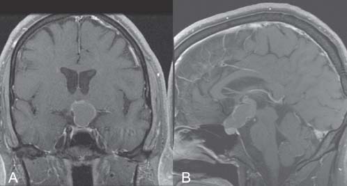
Case 13 Craniopharyngioma: Endoscopic Approach


Daniel M. Prevedello, Amin B. Kassam, Paul Gardner, Arlan H. Mintz, Carl H. Snyderman, and Ricardo L. Carrau


Image

Fig. 13.1 (A) Coronal and (B) sagittal T1-weighted magnetic resonance images of the brain with contrast showing a suprasellar mass.


Image Clinical Presentation


Image Questions


  1. What is the diagnosis?
  2. What is the differential diagnosis?
  3. What are the treatment options?
  4. What are the classic surgical approaches for craniopharyngiomas and their main limitations?
  5. How are craniopharyngiomas classified in relation to the infundibulum? Which type is demonstrated in this case?

    You decide to approach the tumor via an endoscopic endonasal route.


  6. Describe the advantages of an endoscopic endonasal approach (EEA) over the standard transcranial approaches.
  7. What are the limitations for EEAs for the treatment of craniopharyngiomas?
  8. What are the prognostic factors for risks of recurrence or regrowth?

Image Answers


  1. What is the diagnosis?

    • Craniopharyngioma is most likely – The diagnosis is made based on both radiologic and clinical findings.
    • The contrast-enhanced MRI demonstrates a heterogeneous enhancing sellar and suprasellar lesion with solid and cystic components.
    • In addition, CT images confirm the presence of calcifications in the lesion, which are present in ~80% of craniopharyngioma cases.
    • Clinically, the presentation of panhypopituitarism and occasionally diabetes insipidus combined with these imaging studies is diagnostic for craniopharyngioma.

  2. What is the differential diagnosis?

  3. What are the treatment options?

    • The main treatment modality for craniopharyngiomas is surgical resection. A total surgical resection decreases the possibility of a tumor recurrence. The first surgical attempt is the most important in determining the outcome because reoperation on a recurrent or residual craniopharyngioma is less likely to result in a complete resection due to scar tissue formation caused by the previous surgery or radiation.2
    • Other modalities of treatment, in general reserved for recurrent tumors, include

      • Radiation (radiosurgery, intensity modulated radiation therapy, or fractionated radiotherapy)2,3
      • Stereotactic cystic drainage (with or without Ommaya reservoir placement)
      • Stereotactic intracystic delivery of radioactive or immunoactive substances

        • Bleomycin4
        • Radioactive substances

          • Phosphorous-32 (P32)5
          • Yttrium-90 (Y90)6
          • Rhenium-186 (Re186)7

        • Interferon-α8

  4. What are the classic surgical approaches for craniopharyngiomas?

    • Frontal interhemispheric and pterional transsylvian approaches9:

      • May include orbitozygomatic osteotomies and other variations
      • The cranial–caudal angle of approach has a substantial limitation with the optic apparatus positioned between the surgeon and the target.
      • To improve corridors around the parachiasmatic space, critical positioning of the chiasm needs to be considered via these approaches, specifically whether the chiasm is positioned anteriorly (prefixed) or posteriorly (postfixed).
      • Anterior interhemispheric approach with subsequent opening of the lamina terminalis

        • This is an option for tumors eroding the floor of the 3rd ventricle. It is a midline approach well indicated to resect a midline lesion; however, it is very limited in the exposure of the undersurface of the optic apparatus and is usually indicated only for intraventricular craniopharyngiomas.

    • Lateral presigmoid combined with a transtentorial subtemporal approach

      • Option to minimize optic apparatus manipulation is a caudal–cranial angle of attack.
      • Allows for exposure of the pre-pontine and interpeduncular cisterns.
      • Nevertheless, any dissection through this lateral view is divided into many small corridors in between the cranial nerves (II to XII) that are present in the midline cisterns’ lateral walls.

  5. How are craniopharyngiomas classified in relation to the infundibulum? Which type is demonstrated in this case?

    • The classification is summarized in Fig. 13.2.10

      • Type I is preinfundibular.
      • Type II is transinfundibular (extending into the stalk) – This case illustration.
      • Type III is retroinfundibular (extends behind the gland and stalk).11
      • Type IV is isolated to the 3rd ventricle and/or optic recess and may not be accessible via an endonasal approach.

  6. Describe the advantages of an EEA over the standard transcranial approaches.

  7. What are the limitations for EEA for the treatment of craniopharyngiomas?

    • Lack of space and/or three-dimensional perception

      • Freedom of movements is improved by using two surgeons synergistically performing the surgery through both nostrils coupled with better depth perception generated by continuous movements of the camera.

    • Any extension of the lesion laterally beyond the optic nerves formally determines a limitation for an endoscopic endonasal resection of craniopharyngiomas. Lesions located in those territories have to be accessed through paramedian corridors (craniotomies) either as a complementary or an exclusive approach for resection of specific lesions.
    • Lack of surgeon’s experience with the endoscope.12

  8. What are the prognostic factors for risks of recurrence or regrowth?

    • Favorable prognostic factors for risk of recurrence/regrowth in craniopharyngioma include2

      • Size (<4 cm)
      • Favorable location
      • Complete surgical removal
      • Age (>5 years carries favorable prognosis)
      • Absence of severe hypothalamic involvement
< div class='tao-gold-member'>

Only gold members can continue reading. Log In or Register to continue

Stay updated, free articles. Join our Telegram channel

Jul 16, 2016 | Posted by in NEUROSURGERY | Comments Off on 13 Craniopharyngioma: Endoscopic Approach

Full access? Get Clinical Tree

Get Clinical Tree app for offline access