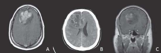
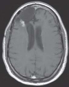
Case 14 High-grade Glioma: Surgical Treatment Fig. 14.1 T1-weighted magnetic resonance images (MRIs) of the head with contrast enhancement, axial (A) and coronal (C) sections. (B) Computed tomography (CT) scan of the head with contrast enhancement. Fig. 14.2 T1-weighted magnetic resonance image (MRI) of the head with contrast enhancement, axial section, 10 months postresection.
 Clinical Presentation
 Questions
 Answers
< div class='tao-gold-member'>
Only gold members can continue reading. Log In or Register to continue


