16 Eloquent Cortex Low-grade Glioma

Case 16 Eloquent Cortex Low-grade Glioma


John Winestone and Allen K. Sills Jr.


Image

Fig. 16.1 (A) T1- and (B) T2-weighted axial magnetic resonance images of the brain at the level of the temporal lobe.


Image Clinical Presentation


Image Questions


  1. Describe the images. (Note: The lesion is nonenhancing on contrasted images – not shown here.)
  2. What is the differential diagnosis?
  3. What is it about the nature of her seizures that can help to localize them?
  4. Which hemisphere would you expect to be dominant and how may it be determined?
  5. What is a Wada test and how would it assist in planning treatment?
  6. Describe any further investigation and how it would guide you in your treatment plans.
  7. What is the treatment for diffuse low-grade glioma in eloquent cortex?
  8. Describe some adjuncts used in surgical resection.

    The patient underwent surgical resection of the mass via a right temporal approach. The sylvian fissure was split, and a corridor to the lesion was opened via a corticotomy in the mesial temporal lobe. Abnormal tissue was identified and confirmed by a pathologist. Tumor was removed until clear margins were reached in all planes. The pathology was consistent with low-grade oligodendroglioma.


  9. What should the follow-up of this patient include?
  10. What does the 1p 19q genetic mutation imply?
  11. What is the prognosis?


Image

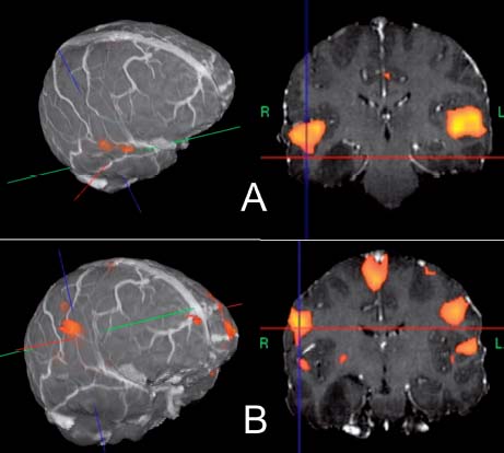
Fig. 16.2 Functional magnetic resonance imaging (fMRI) demonstrating bilaterality of (A) listening and (B) speech areas in the brain.
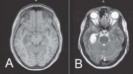

Image Answers


  1. Describe the images.

    • There is a 3 × 2-cm temporal mass on the right side, located in the mesial temporal lobe.
    • The mass is hypointense on T1- and hyperintense on T2-weighted images.
    • There is no significant mass effect or midline shift.

  2. What is the differential diagnosis?

    • The differential diagnosis includes1

      • Low-grade glioma
      • Lymphoma
      • Demyelinating lesion
      • Infectious pathologies (such as herpes encephalitis)
      • Other less likely possibilities include metastases, infarction, arachnoid cyst, epidermoid.

  3. What is it about the nature of her seizures that can help to localize them?

    • Her neurologic symptoms describe a partial complex seizure, which may be traced to the temporal lobe.

  4. Which hemisphere would you expect to be dominant and how may it be determined?

    • The presence of a right-sided lesion causing language-related problems is suggestive of bihemispheric language input.

  5. What is a Wada test and how would it assist in planning treatment?

    • A Wada test (intracarotid sodium amobarbital procedure) consists of a selective intracarotid amobarbital injection while performing an angiogram.2
    • Neuropsychological testing is performed on an awake patient, and deficits are observed in speech and memory as hemiplegia occurs on the side of injection of amobarbital.
    • A Wada test can help localize speech when there is concern for bihemispheric input.
    • It is also used to assess temporal lobe dominance in terms of memory.

  6. Describe any further investigation and how it would guide you in your treatment plans.

    • Functional MRI (fMRI), electroencephalogram, positron emission tomography, or single photon emitted computed tomography scan2,3
    • The fMRI (Fig. 16.2) clearly demonstrates speech areas away from the lesion, even on the ipsilateral side.
    • This finding enables planning of surgical resection with the possibility of sparing of eloquent cortex.

  7. What is the treatment for diffuse low-grade glioma in eloquent cortex?

  8. Describe some adjuncts used in surgical resection.

    • Image guidance is a useful instrument in resection. Its reliability must be tempered with the inherent limitations of most guidance systems.
    • Intraoperative imaging
    • Frameless navigation
    • Ultrasound, mapping
    • Awake craniotomy
    • Direct cortical electrocorticography3

  9. What should the follow-up of this patient consist of?

    • National Comprehensive Cancer Network (NCCN) guidelines recommend maximal resection where possible, followed by observation in younger patients. In those older than 45, external-beam radiation and/or chemotherapy in tumors with 1p/19q deletions are recommended. Patients should be followed with annual MRIs indefinitely.6

  10. What does the 1p 19q genetic mutation imply?

    • More likely to be of oligodendroglial origin7
    • Better prognosis8
    • More responsive to chemotherapy8
    • Longer tumor-free survival after chemotherapy8
    • Chemotherapy used for treatment may include PCV (procarbazine, CCNU, vincristine) with 50% complete resolution versus non 1p-19q → only 25% response.7
    • Temozolomide may be used for recurrent tumors.8

  11. What is the prognosis?

    • Mean survival = 4.4–9.8 years (after operative treatment)5,7
    • 5-year survival = 38–75%
    • 10-year survival = 19–59%
< div class='tao-gold-member'>

Only gold members can continue reading. Log In or Register to continue

Stay updated, free articles. Join our Telegram channel

Jul 16, 2016 | Posted by in NEUROSURGERY | Comments Off on 16 Eloquent Cortex Low-grade Glioma

Full access? Get Clinical Tree

Get Clinical Tree app for offline access