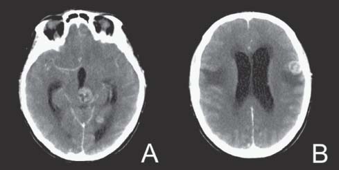
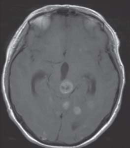
Case 18 Multiple Brain Metastases Fig. 18.1 (A,B) Computed tomography (CT) scans of the head with contrast brain windows. Fig. 18.2 T1-weighted magnetic resonance image (MRI) of the brain with contrast, axial section.
 Clinical Presentation
 Questions
 Answers
< div class='tao-gold-member'>
Only gold members can continue reading. Log In or Register to continue


