20 Primary Central Nervous System Lymphoma

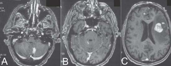
Case 20 Primary Central Nervous System Lymphoma


John Winestone and Allen K. Sills Jr.


image

Fig. 20.1 (A–C) Enhanced T1-weighted magnetic resonance images showing multiple lesions, (C) the largest one being in the left frontal lobe.


Image Clinical Presentation


Image Questions


  1. Describe the MRI.
  2. What is the differential diagnosis for the imaging findings in this scenario?
  3. What is an appropriate workup to help narrow the differential diagnosis?
  4. If a tissue sample was needed, which would be the best lesion to biopsy?

    After a metastatic and immunodeficiency workup was negative, we elected to biopsy the left frontal lobe lesion. Care was taken to have the patient off of steroids for 2 weeks before the biopsy. The pathology results confirmed central nervous system (CNS) lymphoma.


  5. Name the different ways to diagnose CNS lymphoma.
  6. Describe the treatment course.
  7. What is the role of surgery?
  8. What is the outcome with and without treatment?

Image Answers


  1. Describe the MRI.

    • The MRI shows multiple enhancing lesions. They are dispersed throughout the brain with some lesions abutting the ventricular system.
    • The largest lesion is in the left frontal lobe, measuring ×2 Ø 2.5 cm with some local edema.
    • The lesions are bilateral.

  2. What is the differential diagnosis for the imaging findings in this scenario?

  3. What is an appropriate workup to help narrow the differential diagnosis?

    • An aggressive medical workup searching for neoplastic disease is in order. CT scans of the viscera as well as basic screening tests for common malignancies such as breast, lung, prostate, and colon cancer would help define a metastatic source.
    • Blood work for neoplastic markers as well as immunodeficiency (prostate-specific antigen, human immunodeficiency virus, carcinoembryonic antigen) may aid in the workup.
    • Cerebrospinal fluid (CSF) sampling may be helpful in determining demyelinating disease as well as detecting cells from a lymphoma process including tumor markers such as lactate dehydrogenase.
    • Beta-glucuronidase and beta 2-microglobulin may also be sought out in the CSF.2,3

  4. If a tissue sample were needed, which would be the best lesion to biopsy?

    • The left frontal lesion is the most easily accessible for tissue diagnosis. If there were complications from the surgery, this location would be the most tolerant in terms of subsequent deficits.

  5. Name the different ways to diagnose CNS lymphoma.

    • National Comprehensive Cancer Network (NCCN) guidelines recommend holding steroids when lymphoma is suspected by history, physical examination, and imaging.
    • The diagnosis can be made with CSF cytology and flow cytometry, a biopsy of the lesion, and/or ophthalmologic examination.
    • The lesion having dissipated with prior steroids is suggestive of PCNSL and a trial of steroids followed by biopsy of any growing lesion is the treatment of choice. Almost all patients will develop resistance after prolonged exposure to steroids. Consequently, steroids should be avoided in suspected but undiagnosed PCNSL. Steroid-induced remission may impact biopsy results2
    • Systemic workup for the hematologic or solid organ source of the lymphoma is suggested.3

  6. Describe the treatment course.

    • High-dose methotrexate is the treatment of choice. Intrathecal methotrexate should be used when there is evidence of disease in the CSF by lumbar puncture or on a MRI of the spine.2,3
    • Radiation therapy is currently not recommended for PCNSL when the patient is older than 60 years.2,3
    • Low-dose whole brain radiation treatment may be used as an adjunct to chemotherapy in selective elderly patients.2,4

  7. What is the role of surgery?

    • Surgery does not improve survival and has a limited role.2

      • Masses large enough to cause impending herniation
      • For diagnostic purposes

  8. What is the outcome with and without treatment?

    • Survival

      • Median survival less than 3 months without treatment (supportive care only)5
      • Median overall survival is 37 months with treatment.6
      • Median survival in elderly patients is 7 months (including partial or limited treatment).4

    • Predictors of poor survival include increased age, low Karnofsky performance score, hemiparesis, altered mental status, and decreased creatinine clearance.6
< div class='tao-gold-member'>

Only gold members can continue reading. Log In or Register to continue

Stay updated, free articles. Join our Telegram channel

Jul 16, 2016 | Posted by in NEUROSURGERY | Comments Off on 20 Primary Central Nervous System Lymphoma

Full access? Get Clinical Tree

Get Clinical Tree app for offline access