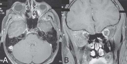
Case 22 Fibrous Dysplasia of the Skull Fig. 22.1 (A) T1-weighted postcontrast FatSat (fat saturation) axial and (B) coronal magnetic resonance images. Note the hyperostotic abnormal bone involving the orbital roof, the posterolateral wall of the orbit, and extending into the infratemporal fossa.
 Clinical Presentation
 Questions
![]()
Stay updated, free articles. Join our Telegram channel

Full access? Get Clinical Tree


