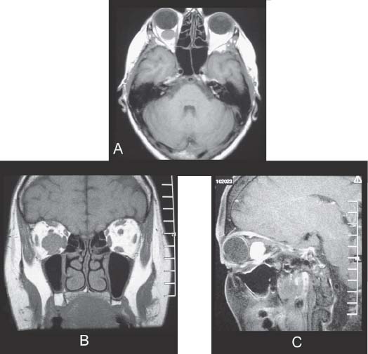
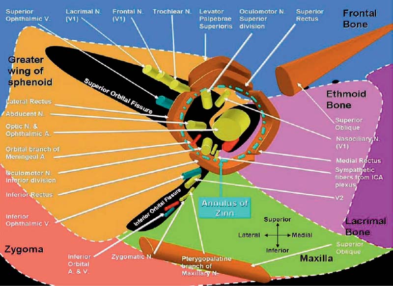
23 Orbital Tumor

Case 23 Orbital Tumor


Michel Lacroix


Image

Fig. 23.1 (A) Axial T1-weighted, (B) coronal T1-weighted, and (C) sagittal T1-weighted magnetic resonance images (MRIs) of the brain with gadolinium contrast brought by the patient during the initial visit.


Image Clinical Presentation


Image Questions


  1. Interpret the MRI.
  2. Give a general classification of orbital tumors.
  3. What structures are contained in the annulus of Zinn?
  4. Give a differential diagnosis.
  5. What is your initial management?
  6. Describe the surgical approach and choose the one you consider best.

    You proceed with a lateral microsurgical approach, achieve a complete macroscopic resection, and the diagnosis is hemangioma.


  7. What is the prognosis and what will be your follow-up?


Fig. 23.2 Artist’s rendering of the content of the annulus of Zinn.


Image Answers


  1. Interpret the MRI.

    • In the right orbit there is a round lesion 1.3 cm in diameter. The lesion is homogeneous and is enhanced strongly after gadolinium injection.
    • The lesion lies inferiorly to the optic nerve to which it may or may not be attached, pushing it upward. There is a significant mass effect with displacement and/or invasion of the inferior rectus muscle and significant secondary proptosis.

  2. Give a general classification of orbital tumors.

  3. What structures are contained in the annulus of Zinn?

    • Cranial nerve (CN) II, ophthalmic artery, CN III (superior and inferior divisions), CN V1 (nasociliary), CN VI (abducens)
    • Figure 23.2

  4. Give a differential diagnosis.

    • The lesion appears to be extraaxial and intraconal and does not involve the eye globe. Considering the appearance of the lesion and the broad differential diagnosis of orbital lesions,2 one should probably list these pathologies in this sequence:

      • Hemangioma: the most common benign lesion of this location
      • Meningioma of the optic nerve sheath
      • Neurofibroma
      • Melanoma: the most frequent primary malignancy in adults in this location or other metastatic tumor
      • Lymphoma: a frequent cause of painless proptosis
      • Vascular, endocrine, infectious, and inflammatory diseases are unlikely. It is not a congenital malformation.

  5. What is your initial management?

  6. Describe the surgical approach and choose the one you consider best.

    • Relying on the MRI, the lesion can be located inferior to the optic nerve and lateral to the optic apex.
    • A lateral microsurgical approach is a viable surgical option4,5 : the skin incision is made superolateral to the eyebrow and is carried posteriorly ~4 cm.
    • A lateral orbito-zygomatectomy allows exposure of the periorbital fascia. A traction suture can be placed to identify the lateral rectus muscle and follows it. The incision in the periorbita is inferior to the lateral rectus muscle.
    • A medial rotation of the globe is performed by light traction. Access is then achieved to the intraconal lesion.
    • For lesions with intracranial extension, lesions involving the optic canal and lesions medial to the optic apex, a transcranial fronto-orbital temporal approach is essential.
    • A medial orbitotomy is preferred for a tumor located anteriorly to the orbit and medial to the optic nerve.

  7. What is the prognosis and what will be your follow-up?

    • A complete resection of a hemangioma is curative. No other treatment or long-term follow-up is required.6
< div class='tao-gold-member'>

Only gold members can continue reading. Log In or Register to continue

Stay updated, free articles. Join our Telegram channel

Jul 16, 2016 | Posted by in NEUROSURGERY | Comments Off on 23 Orbital Tumor

Full access? Get Clinical Tree

Get Clinical Tree app for offline access