24 Multiple Ring-enhancing Cerebral Lesions

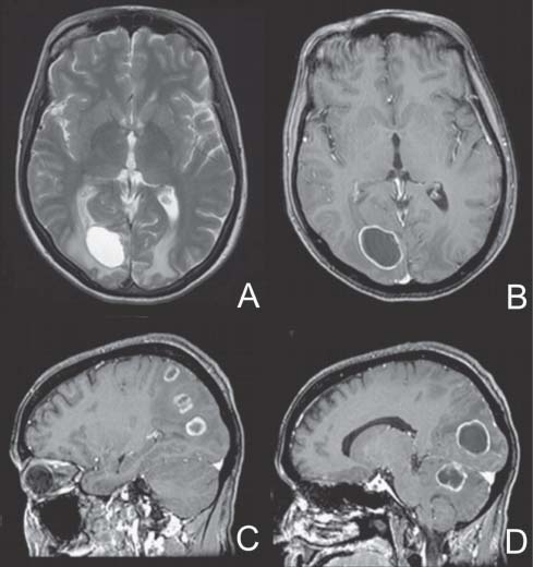
Case 24 Multiple Ring-enhancing Cerebral Lesions


Kevin Petrecca and Rolando Del Maestro


Image

Fig. 24.1 (A) T2-weighted axial magnetic resonance image (MRI), (B) T1-weighted axial MRI with contrast, and (C,D) T1-weighted sagittal MRI showing intraparenchymal lesions.


Image Clinical Presentation


  • A 44-year-old woman presents with sudden onset right ear pain 2 months ago associated with headache, dizziness, and difficulty getting out of bed while vacation ing in the Dominican Republic.
  • Patient is a smoker.
  • She was given a 1-week course of antibiotics for a presumed otitis media.
  • She developed a progressive unsteady gait.

Image Questions


  1. Describe the magnetic resonance imaging (MRI) findings (Fig. 24.1).
  2. What information should be obtained on history?
  3. What is the differential diagnosis?
  4. What are your initial investigations and how would you manage this patient?
  5. Are there any MRI findings that can distinguish a bacterial abscess from a neoplasm?
  6. What are the histologic stages of bacterial abscess formation and what are their radiologic correlates?
  7. What are the sources of bacterial abscesses?

    A stereotactic biopsy for diagnosis was performed and was nondiagnostic and the lesions are not decreasing in size on broad spectrum antibiotics.


  8. What is the next step in your management?

    A right parietal craniotomy was performed and one of the lesions was resected. Pathology was consistent with an adenocarcinoma. A computed tomography (CT) scan of the chest and abdomen revealed a small mass adjacent to the right main stem bronchus.


Image Answers


  1. Describe the MRI findings (Fig. 24.1).

    • The T2-weighted MRI shows a right occipital cystic lesion with surrounding edema. There is also edema in the contralateral occipital lobe. The lesion exhibits a small amount of mass effect, evident as a loss of sulcation in the area of the lesion.
    • The contrast-infused images reveal multiple lesions, a larger lesion in the cerebellar vermis, and two other lesions in the right parieto-occipital region. Each lesion has similar characteristics including a well-formed enhancing capsule surrounding a cystic center. A developmental venous anomaly is present in the left frontal lobe.

  2. What information should be obtained in the history?

    • It is important to identify risk factors for cerebral abscess formation and for metastatic disease: smoking, weight loss, night sweats, fatigue, cough, intravenous drug use, cardiac or pulmonary abnormalities, human immunodeficiency virus status, other high-risk behaviors, etc.

  3. What is the differential diagnosis?

    • The differential diagnosis includes multiple bacterial abscesses, neurocysticercosis, toxoplasmosis, tuberculomas, metastases, and multifocal glioblastoma.

  4. What are your initial investigations and how would you manage this patient?

    • Markers of infections include an elevated white blood cell count, erythrocyte sedimentation rate, and C-reactive protein.
    • A chest radiograph to rule out pneumonia, tuberculosis, and a neoplasm
    • Metastatic workup may be initiated with CT of the chest, abdomen, and pelvis.
    • Gram stain, blood and urine cultures to identify a hematogenous origin to an abscess as well as serum antitoxoplasma titers
    • Radiographs of the arms and legs to identify subcutaneous or muscular calcifications that can be present in cysticercosis
    • Western blot tests to detect Taenium solium antigens from serum
    • An electrocardiogram to assess cardiac rhythm and an abdominal ultrasound to identify a mass

  5. Are there any MRI findings that can distinguish a bacterial abscess from a neoplasm?

    • Cerebral abscesses can mimic necrotic tumors and cystic metastases on conventional MRI. These lesions can often, however, be differentiated on diffusion-weighted imaging (DWI).
    • Cerebral abscesses typically demonstrate restricted diffusion on DWI, whereas tumors and cystic metastases usually do not.1

  6. What are the histologic stages of bacterial abscess formation and what are their radiologic correlates?

    • There are four histologic stages in cerebral abscess formation2,3:

      • Early suppurative cerebritis (days 1–2) defined by endothelial cell swelling, perivascular neutrophil infiltration. CT findings include an area of hypodensity that may exhibit patchy enhancement.
      • Late suppurative cerebritis with confluent central necrosis (days 3–7) defined by adjacent foci of necrosis, which enlarge and become confluent. The infiltrate now includes macrophages, lymphocytes, and plasma cells. CT findings include a more pronounced central hypodensity with a thick enhancing ring surrounded by a hypodensity.
      • Early encapsulation (days 8–14) defined by capsular neovascularity, fibroblast infiltration, collagen deposition, and perilesional edema. CT findings include a well-developed central core with a thinner well-formed enhancing ring.
      • Late encapsulation (days >14) defined by central necrosis, a thin collagen capsule and lymphocytes. Note: Capsule is thinner along the ventricular wall, increasing the susceptibility of rupture into ventricular system. CT findings include a very thin enhancing ring surrounded by a hypodensity.

  7. What are the sources of bacterial abscesses?

    • There are three sources of cerebral bacterial abscesses.
    • The most common route is hematogenous, which accounts for 25% of abscesses. The most common pathogen is Streptococcus viridans.
    • A second etiology is from a contiguous source such as a paranasal sinus, middle ear, dental root, osteomyelitis, or emissary vein. The most common pathogen is Streptococcus milleri.
    • The third route is direct from a trauma or postsurgical, especially postsinus breach. The common pathogens are Staphylococcus aureus and epidermidis.3

  8. What is the next step in your management?

    • Because the stereotactic biopsy was nondiagnostic and the lesions are not decreasing in size on broad-spectrum antibiotics, a more invasive approach to make a diagnosis is necessary.
    • Treatment options include steroid therapy for symptom relief, resection of one or more lesions followed by whole brain radiotherapy (WBRT), radiosurgery for each lesion with or without WBRT, or WBRT alone.
< div class='tao-gold-member'>

Only gold members can continue reading. Log In or Register to continue

Stay updated, free articles. Join our Telegram channel

Jul 16, 2016 | Posted by in NEUROSURGERY | Comments Off on 24 Multiple Ring-enhancing Cerebral Lesions

Full access? Get Clinical Tree

Get Clinical Tree app for offline access