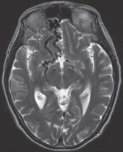
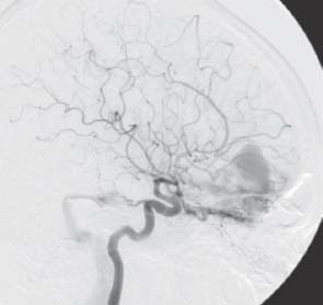
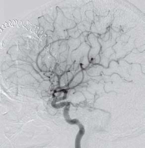
27 Dural Arteriovenous Fistula

Case 27 Dural Arteriovenous Fistula


Nancy McLaughlin and Michel W. Bojanowski


Image

Fig. 27.1 Axial T2-weighted magnetic resonance image of the head.


Image

Fig. 27.2 Digital subtraction angiography lateral view, right carotid injection.


Image Clinical Presentation


  • A 68-year-old woman was referred for evaluation of a pulsatile tinnitus ongoing for the past 7 years and a more recent headache of variable intensity occurring daily for one year. The patient is in otherwise excellent health. No other symptoms were reported.
  • Neurologic examination was normal; specifically, no bruit was auscultated over the skull and no pulsatile tinnitus was documented.

Image Questions


  1. What is your differential diagnosis for pulsatile tinnitus?
  2. What are the indications to investigate a pulsatile tinnitus?
    Findings on the magnetic resonance imaging (MRI) scan (
    Fig. 27.1) revealed a vascular anomaly and required an angiography (Fig. 27.2).
  3. Describe the angiography.
  4. What is your management? Justify.
    You decided to treat the anterior cranial fossa dural arteriovenous fistula (DAVF) surgically.
  5. What is the main step during surgery to eliminate the fistula?
  6. What are the possible complications of surgical treatment?
    The postoperative angiography shows a complete exclusion of the anterior cranial fossa DAVF. However, there is now a retrograde venous drainage from the sigmoid sinus fistula (Fig. 27.3).
  7. What is your management?
  8. What are the causes of DAVFs?
  9. How are DAVFs classified?


Image

Fig. 27.3 Digital subtraction angiography postoperative lateral view, right carotid injection.


Image Answers


  1. What is your differential diagnosis for pulsatile tinnitus?

  2. What are the indications to investigate a pulsatile tinnitus?

    • In the absence of an audible bruit, MRI–magnetic resonance angiography (MRA) is an appropriate initial diagnostic step for subjective pulsatile tinnitus.
    • In the presence of an objective pulsatile tinnitus, the clinician may proceed initially with an MRI–MRA. However, in patients with an audible bruit and those with a history of trauma accompanied with de novo pulsatile tinnitus, an angiography is warranted.1

  3. Describe the angiography.

    • The angiography showed a right anterior cranial fossa DAVF supplied by branches of the right and left ophthalmic arteries, right and left internal maxillary arteries, and right middle meningeal artery.
    • A large venous pouch located in the right anterior fossa was identified. Retrograde venous drainage to cortical veins toward the basal vein of Rosenthal and great cerebral vein of Galen was present.
    • Other cortical veins refluxed toward the longitudinal superior sagittal sinus.
    • The angiography also revealed a right sigmoid sinus fistula nourished by the right tentorial artery. No retrograde venous reflux was noted (Fig. 27.2).

  4. What is your management? Justify.

    • Treatment is indicated to eliminate the risk of hemorrhages and neurologic deficits. The overall morbidity and mortality rate of patients harboring a DAVF with cortical venous retrograde drainage (CVR) are 15% and 10%, respectively.3
    • Anterior cranial fossa DAVFs always drain via cortical venous drainage and therefore mandate an aggressive treatment.4
    • Transarterial embolization is limited to branches of the external carotid artery. Embolization of ophthalmic arteries is avoided due to the inherent risk of central retinal artery occlusion.
    • Transvenous embolization is not feasible for most of these fistulas because of the lack of venous access.
    • Surgery is the best treatment option for anterior cranial fossa dural arteriovenous fistulas.5,6
    • Lesions without CVR such as the sigmoid sinus fistula do not require treatment except if symptoms are intolerable. In such situations, a palliative treatment may be indicated.4

  5. What is the main step during surgery to eliminate the fistula?

    • The goal is to interrupt the draining veins at their dural origin.

  6. What are the possible complications of surgical treatment?

    • Possible complications of anterior fossa DAVFs treated by surgical means include excessive blood loss, venous hypertension, venous infarct, cerebral edema, and seizures.

  7. What is your management?

    • Prior to surgery of the anterior fossa DAVF, the sigmoid fistula was initially considered for conservative treatment.
    • However, after the surgery of the anterior fossa DAVF, the sigmoid fistula developed a retrograde venous drainage and a curative treatment for this fistula should be sought.
    • Transvenous embolization should be considered initially.
    • If endovascular treatment fails, surgery should be performed for patients with CVR.
    • Whenever possible, transarterial embolization should be attempted to reduce int raoperative bleeding.4

  8. What are the causes of DAVFs?

    • The etiology of DAVFs remains poorly understood. Some conditions have been associated with DAVF such as head injury, prior craniotomy, infection, arterial dysplasia, dural venous sinus thrombosis.
    • Most of these etiologies have a sinus occlusion in common.

  9. How are DAVFs classified?
< div class='tao-gold-member'>

Only gold members can continue reading. Log In or Register to continue

Stay updated, free articles. Join our Telegram channel

Jul 16, 2016 | Posted by in NEUROSURGERY | Comments Off on 27 Dural Arteriovenous Fistula

Full access? Get Clinical Tree

Get Clinical Tree app for offline access