28 Cerebral Arteriovenous Malformation

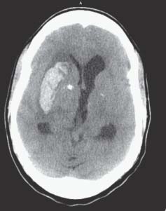
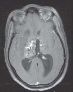
Case 28 Cerebral Arteriovenous Malformation


Pascal M. Jabbour and Erol Veznedaroglu


Image

Fig. 28.1 Computed tomography scan of the head showing a right basal ganglia bleed.


Image

Fig. 28.2 Magnetic resonance imaging scan of the head showing a basal ganglia arteriovenous malformation.


Image Clinical Presentation


Image Questions


Image Answers


  1. Considering the age of the patient, what is the most likely etiology of the bleed?

    • Any intracerebral hemorrhage in a young patient without any past medical history should raise the suspicion of a vascular malformation.

  2. What is the next test to order?

    • The CT of the head showed a basal ganglia bleed in a young patient; the next test to be ordered should be an imaging modality that is able to demonstrate any vascular abnormality, like a CT angiogram, MRI, or magnetic resonance angiography.

  3. What is the most common AVM grading system used? And what is the patient’s grade?

  4. What is the risk of rupture of an AVM and the lifetime cumulative risk for this patient?

    • The risk of rupture of this AVM is ~4% per year.2
    • Risk is 1 (risk of not hemorrhaging) raised to the power of years left to live.
    • In a 36-year-old patient, at an average life expectancy of 79 years, the years left to live can be calculated to be 43 years.
    • Risk = 1 – (1 – risk of hemorrhage) 43
    • Risk = 1 – (0.96) 43 = 83
    • The patient has a lifetime risk of hemorrhage of 83%.

  5. What are the diff erent treatment options for this patient?

    • The diff erent options are1,3,4

      • No intervention if the risk of intervening is higher than the natural history risk of the AVM rupturing.
      • Embolization in preparation for surgical resection
      • Embolization with the goal of reducing the volume of the AVM in preparation for radiosurgery
      • Surgery alone
      • Radiosurgery alone with possible volume fractionation

  6. What are some important steps of AVM surgery?

    • Furosemide + mannitol prior to dural opening
    • Proximal temporary occlusion can be performed with aneurysm clips.
    • Smaller vessels are occluded with AVM mini-clips.
    • Large draining veins are clipped with large aneurysm clips.
    • Do not commit on vessel occlusion until the vessel is seen to enter the AVM.
    • “If it looks like it may be AVM, it probably is.”
    • Difficult-to-control bleeding during this dissection is commonly an indication that the AVM–brain interface has been breached on the side of the AVM.
    • As a safety measure, before taking the major draining vein, an aneurysm clip is placed across it for 10–15 minutes and dissect out in a diff erent area.
    • Always obtain a postoperative angiogram and CT scan.5

  7. What is normal perfusion pressure breakthrough and how is it treated?

    • It is a disorder of autoregulation of the brain vasculature.
    • May present with sudden onset of brain swelling and bleeding from multiple sites
    • May be due to draining vein that was taken too soon
    • Also occurs from raw normal brain surfaces, that have lost autoregulatory capacity
    • Close inspection of the operative field will identify any residual AVM.
    • Treatment

      • Elevation of the patient’s head
      • Administration of mannitol and furosemide
      • Barbiturate- or etomidate-induced coma
      • Hypotension may be beneficial.
      • Focal hypotension by means of proximal vessel occlusion with temporary clip5
< div class='tao-gold-member'>

Only gold members can continue reading. Log In or Register to continue

Stay updated, free articles. Join our Telegram channel

Jul 16, 2016 | Posted by in NEUROSURGERY | Comments Off on 28 Cerebral Arteriovenous Malformation

Full access? Get Clinical Tree

Get Clinical Tree app for offline access