29 Cavernous Angioma

Case 29 Cavernous Angioma


Julius July and Eka Julianta Wahjoepramono


Image

Fig. 29.1 T2-weighted magnetic resonance image showing a lesion involving the right basal ganglia and thalamus.


Image 
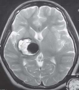
Clinical Presentation


Image Questions


  1. Describe the MRI feature and provide a differential diagnosis.
  2. What are the treatment options?
  3. What treatment would you recommend for this case?
  4. What is your argument between a surgical and nonsurgical option?
  5. If you decide to do the surgery, what is your approach to remove the lesion?
  6. What are the potential complications of surgery?

Image Answers


  1. Describe the MRI feature and provide a differential diagnosis.

  2. What are the treatment options?

    • Conservative treatment

      • Asymptomatic or minimally symptomatic lesions because they may remain quiescent and if they rebleed, this tends to be mild and not catastrophic.
      • Multiple lesions with familial history
      • Deep or eloquent area when the surgical risks exceed the benefit, especially in elderly patients

    • Radiosurgery

      • In cases of surgically inaccessible lesions with at least one prior hemorrhage
      • Several studies show that radiosurgery could reduce the risk of hemorrhage to one-third of initial risk.
      • However, because of the high complication rate of radiosurgery, its routine use for cavernoma fell out of favor. The benefits of radiosurgery are difficult to assess because of the unclear natural history of cavernomas, the inability to evaluate the status of the malformation vessels, and the lack of completeness of obliteration of the malformation.

    • Surgery

      • Recurrent hemorrhage, especially at a young age
      • Progressive neurologic deficits
      • Intractable epilepsy when the benefit outweighs the risk
      • Lesion located in the cerebellum or the cerebral cortex
      • Lesions that do not respond to radiosurgery
      • In special circumstances (e.g., a young woman who wants to become pregnant with an accessible lesion)
      • To prevent future bleeding2,3

  3. What treatment would you recommend for this case?

    • Considering the patient’s young age and history of repeated hemorrhage with progressive neurologic deterioration, surgical treatment should be recommended.

  4. What is your argument bet ween a sur gical and nonsurgical option?

    • Kondziolka et al.’s4 prospective study of 122 cavernomas has shown that the annual bleeding rate for symptomatic lesions is 4.5%.
    • The patient has a 93.6% chance of sustaining another hemorrhage in her lifetime (with life expectancy of 79 years, based on formula from Case 28). The hemorrhage could be catastrophic.1
    • Although the lesion is located in eloquent brain (thalamus-basal ganglia), surgical resection still provides significant benefits that are greater than the above risk.
    • Based on the MRI (Fig. 29.1), the sylvian fissure appears to be quite relaxed. Resection can be achieved with image-guided surgery by performing a sylvian fissure dissection. A corticotomy along the insular gyrus of ~1–2 cm is enough to remove the cavernoma (Fig. 29.2 and Fig. 29.3).
    • Alternatively, a parietal transcortical-transventricular approach may also be used.

  5. What are the potential complications of surgery?

    • The complications of surgery consist of general risk that might happen during any neurosurgical cases (such as infection, cerebrospinal fluid leak leak, seizure, stroke, deep vein thrombosis, pneumonia, coma, death, etc.).
    • They also include neurologic worsening related to lesion location (such as paraesthesia, hemiparesis or hemiplegia, visual field deficits, etc.).3,4
< div class='tao-gold-member'>

Only gold members can continue reading. Log In or Register to continue

Stay updated, free articles. Join our Telegram channel

Jul 16, 2016 | Posted by in NEUROSURGERY | Comments Off on 29 Cavernous Angioma

Full access? Get Clinical Tree

Get Clinical Tree app for offline access