31 Unruptured Anterior Communicating Artery Aneurysm

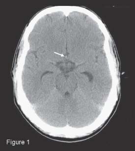
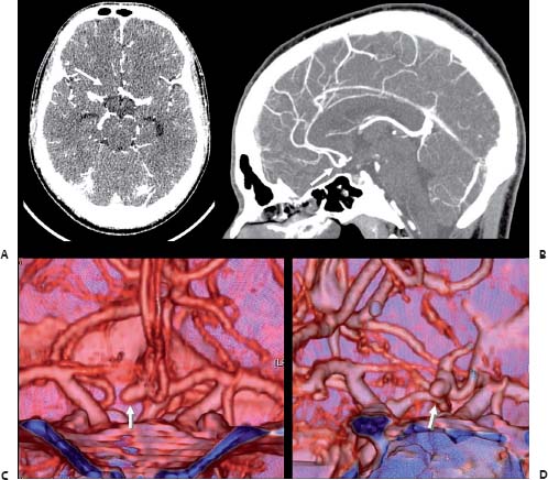
Case 31 Unruptured Anterior Communicating Artery Aneurysm


Yasser I. Orz


Image

Fig. 31.1 Computed tomography scan of the head showing hyperdensity in the interhemispheric fissure (arrow).


Image

Fig. 31.2 (A) Axial and (B) sagittal contrast-enhanced T1-weighted magnetic resonance angiography images, with three-dimensional reconstructions (C) anteroposterior and (D) oblique views.


Image Clinical Presentation


Image Questions


  1. Interpret the CT findings.
  2. What is the most probable diagnosis?
  3. What other investigations would you order?
  4. What are the options and associated risks for management of the lesion?
  5. What is the natural history of unruptured aneurysms?
  6. Name factors favoring treatment of unruptured cerebral aneurysms.
  7. How would you classify anterior communicating artery (ACOM) aneurysms?
  8. If you chose to manage the lesion surgically, what approaches may be utilized?
  9. In the case of a pterional approach, what side would you choose?
  10. In the case of surgery of nonruptured aneurysms, what are less-favorable outcome factors?

Image Answers


  1. Interpret the CT findings.

    • CT of the brain showed a small hyperdense lesion in the suprasellar interhemispheric cistern at the site of the ACOM.

  2. What is the most probable diagnosis?

    • The most probable diagnosis is a nonruptured ACOM aneurysm.

  3. What other investigations would you order?

    • Other investigations (see Case 36 for a detailed comparison of the different treatment measures)
    • Three-dimensional CT angiography (CTA) (Fig. 31.2)

      • It is a noninvasive reliable method for diagnosis of cerebral aneurysms with sensitivity of 95%.
      • In some centers, CTA has become the only diagnostic and pretreatment planning study for patients with ruptured and unruptured cerebral aneurysms.

    • Conventional cerebral angiography

      • The gold standard for evaluation of cerebral aneurysms
      • Four-vessel digital subtraction angiography (DSA) should be done to exclude the presence of multiple aneurysms.

    • Magnetic resonance angiography (MRA)

      • There are several parameters that affect the MRA’s ability to detect the intracranial aneurysms including

        • Aneurysm size
        • Rate and direction of blood flow in the aneurysm relative to the magnetic field

      • Currently MRA may be useful as a screening test in high-risk patients.

  4. What are the options and associated risks for management of the lesion?

    • Occlusion of this aneurysm can be done either by microsurgical clipping (see Fig. 31.3 for a postclipping DSA) or by endovascular coiling.14
    • Alternatively, observation may be an option if the patient so chooses.
    • Table 31.1 summarizes surgical morbidity rates based on size and location.2
    • Table 31.2 summarizes overall risks of coiling of unruptured aneurysms.35

  5. What is the natural history of unruptured aneurysms?

    • According to the International Study of Unruptured Intracranial Aneurysms (ISUIA),2 risk of bleeding from unruptured cerebral aneurysms differs from aneurysms that have ruptured.
    • The natural history and treatment results are affected by the following factors14:

      • Patients’ factors

        • Smoking6
        • Age of the patient
        • Associated medical conditions
        • History of previous subarachnoid hemorrhage (SAH)

      • Aneurysm characteristics

        • Size

          • The most important predictor for future rupture2 as the estimated annual risk of rupture for aneurysms less than 10 mm in diameter is 0.05% and ~1% or more for aneurysms with diameter more than 10 mm, based on initial ISUIA study results of 1998.2
          • ISUIA follow-up study from 20037 stated that 5-year cumulative rupture rates for patients with unruptured aneurysm at internal carotid artery (ICA), ACOM, anterior cerebral artery (ACA), and middle cerebral artery (MCA) are 0%, 2.6%, 14.5%, and 40% for aneurysms less than 7 mm, 7–12 mm, 13–24 mm, and 25 mm or greater, respectively, compared with rates of 2.5%, 14.5%, 18.4%, and 50%, respectively, for the same size categories involving posterior circulation and posterior communicating artery (PCOM) aneurysms.
          • The relative risk is 1.11/mm in diameter.6

      • Location

        • PCOM, vertebrobasilar, and basilar termination unruptured aneurysms are more likely to rupture.

      • Aneurysm morphology: The irregular and multilobular aneurysms are more likely to rupture.

    • Table 31.3 summarizes rupture rates based on aneurysm size.2

  6. Name factors favoring treatment of unruptured cerebral aneurysms.

    • These factors include

      • Young age
      • Previous SAH
      • Larger aneurysm size
      • Aneurysm location and configuration

    • Juvela et al.6 stated that the cumulative rate of unruptured aneurysm rupture is 10.5% at 10 years, 23% at 20 years, and 30.3% at 30 years after diagnosis.

  7. How would you classify ACOM aneurysms?

    • There are two classification schemes of ACOM aneurysms (Fig. 31.4).
    • Yasargil classified the ACOM aneurysms into8

      • Superior
      • Inferior
      • Anterior
      • Posterior

    • Kobayashi classified ACOM aneurysms in terms of approach selection.9

      • Anterior type: (Between 1 and 5 o’clock) The aneurysm lies away from the brain and in relation to the arachnoid membrane. Therefore, wide dissection of the arachnoid lessens the degree of frontal lobe retraction.
      • Inferior type: (Between 5 and 9 o’clock) The aneurysm is likely to be adherent to the chiasm. Therefore, frontal lobe retraction can be dangerous and should be done very carefully.
      • Superior (posterior) type: (Between 9 and 1 o’clock) The aneurysm is usually hidden behind the parent arteries or the gyrus rectus. Therefore, it is difficult to clip via the pterional approach and it may be more suitable to coil them.

  8. If you chose to manage the lesion surgically, what approaches may be used?

    • Surgical approaches that can be used to clip ACOM aneurysms include

      • Pterional approach: This is the most commonly used approach for ACOM aneurysms. It may be supplemented by the cranio-orbital approach in more diffcult to reach or larger aneurysms.
      • Interhemispheric approach: It is infrequently used and indicated if the aneurysm is large or giant and when the aneurysm is in high position (13 mm or more above the anterior clinoid process.8,9

  9. In the case of a pterional approach, what side would you choose?

    • The right pterional approach is the most commonly used approach for the ACOM aneurysms. The left pterional approach is indicated in the following situations:

      • The left A1 is dominant.
      • When the origin of the right A2 is located anterior to that of the left A2, so the neck of the aneurysm is hidden by the right A2 if the right pterional approach is taken.
      • In cases of multiple aneurysms with other aneurysms are located on the left side.

  10. In the case of surgery of unruptured aneurysms, what are less favorable outcome factors?

    • Factors for less favorable outcome post-surgery10:

      • Aneurysm size larger than 15 mm
      • Location: Posterior circulation
      • Number: Multiple aneurysms
      • Associated medical conditions
< div class='tao-gold-member'>

Only gold members can continue reading. Log In or Register to continue

Stay updated, free articles. Join our Telegram channel

Jul 16, 2016 | Posted by in NEUROSURGERY | Comments Off on 31 Unruptured Anterior Communicating Artery Aneurysm

Full access? Get Clinical Tree

Get Clinical Tree app for offline access